Human Biochemistry, Volume 2 - Murray R. 1993
Special Topics
Glycoproteins and Proteoglycans
Proteoglycans and Glycosaminoglycans
Proteoglycans and Glycoproteins are molecules consisting of Proteins with covalently attached oligosaccharide or polysaccharide chains. The distinction between proteoglycans and glycoproteins lies in the Chemical Nature of the Polysaccharides attached to the protein. In proteoglycans, each polysaccharide is composed of repeating disaccharide units that always feature D-glucosamine or D-galactosamine. Each disaccharide component of proteoglycan polysaccharides (with the exception of keratan sulfate) contains a uronic acid—either L-glucuronic acid (GlcUA) or its 5-epimer, L-iduronic acid (IdUA). With the exception of hyaluronic acid, all proteoglycan polysaccharides contain sulfate groups in the form of either O-esters or N-sulfate (in heparin or heparan sulfate).
There are 3 types of linkages between proteoglycan polysaccharides and their polypeptide chain:
1) An O-glycosidic bond between Xyl and Ser, unique to proteoglycans; 2) An O-glycosidic bond between GalNAc and Ser(Thr), present in keratan sulfate II; 3) An N-glycosylamide bond between GlcNAc and the amide nitrogen of Asn.
The pathways of polysaccharide chain formation are completely identical to those involved in the elongation of glycoprotein oligosaccharide chains. UDP-Xyl transferase attaches the Xyl of the sugar nucleotide to Ser, forming a Xyl-Ser O-glycosidic bond. The formation of the O-glycosidic bond between GalNAc and Ser (or Thr) is presumably carried out by a similar UDP-GalNAc transferase. The N-glycosidic bond between GlcNAc and the amide nitrogen of Asn is almost certainly formed with the participation of a lipid-linked polysaccharide, dolichol-P-P-polysaccharide, which, as noted above, is responsible for transporting the preformed oligo- or polysaccharide during glycoprotein synthesis. However, the details of this reaction during proteoglycan synthesis have not yet been elucidated.
The chain elongation process proceeds with the participation of nucleotide sugars acting as Donors. The reactions are regulated primarily by the substrate Specificity of individual Glycosyltransferases. Here again, the "one enzyme, one linkage" principle manifests itself. Reaction specificity depends on the nucleotide sugar donor, the acceptor oligosaccharide, and the anomeric configuration and position of the linkage. The enzymatic systems involved in chain elongation possess The ability to reproduce complex polysaccharides with high precision.
The termination of polysaccharide chain growth results from the following phenomena: 1) the capping effect of sialylation by specific sialyltransferases; 2) sulfation, particularly at the 4-position of the sugars; 3) the removal of the given polysaccharide from the membrane site where catalysis takes place.
Following the Formation of the polysaccharide chain, numerous chemical modifications occur, such as the incorporation of sulfate groups into the GalNAc components of chondroitin sulfate and dermatan sulfate, and the epimerization of the GlcUA residue into an IdUA residue in heparin and heparan sulfate.
An important aspect of proteoglycan METABOLISM is their degradation. Inherited defects in the degradation of proteoglycan polysaccharide chains underlie a group of disorders known as Mucopolysaccharidoses and mucolipidoses, which are discussed below and in Chapter 14. These catabolic disorders have facilitated The Study of specific degradative Enzymes and their substrates. There are A number of exoglycosidases that effect the stepwise Cleavage of sulfate components and glycosyl groups. In addition, there are Endoglycosidases that are present under normal conditions and exhibit varying specificities. For example, the widely distributed enzyme hyaluronidase cleaves N-acetylhexosaminide bonds in hyaluronic acid and chondroitin sulfates.
There are 7 known types of polysaccharides (glycosaminoglycans) that bind to proteins to form proteoglycans. Six of them share a related structure and contain alternating residues of uronic acid and hexosamine within their repeating disaccharide units; keratan sulfate is the exception, as it lacks uronic acid. All of them, except for hyaluronic acid, contain sulfated sugars and are covalently attached to proteins. These 7 types of polysaccharides may differ in their constituent monomers, glycosidic linkages, and the number and Location of sulfate substituents.
Class="center">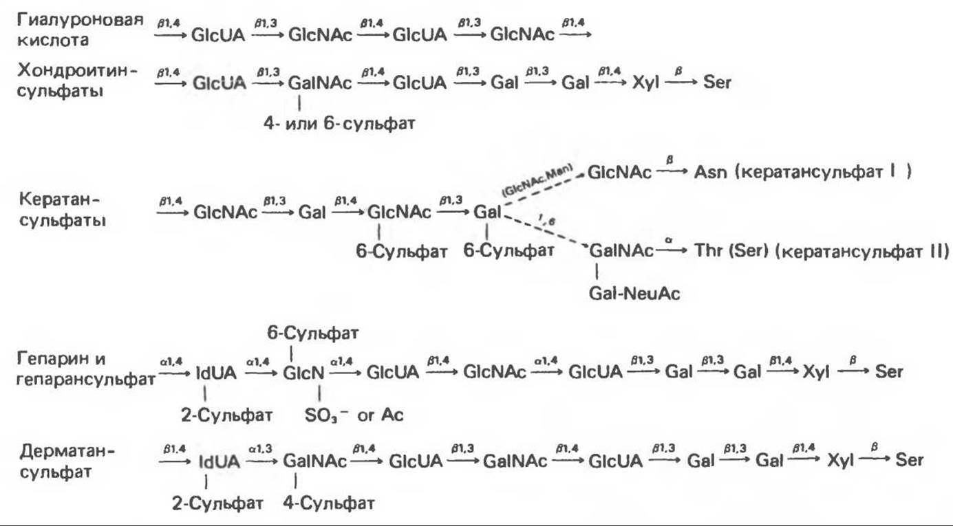
Fig. 54.9. General structure of Proteoglycans and Glycosaminoglycans (GlcUA — D-glucuronic acid; IdUA — L-iduronic acid; GlcN — D-glucosamine; GalN — D-galactosamine; Ac — acetyl; Gal — D-galactose; Xyl — D-xylose; Ser — L-Serine; Thr — L-Threonine; Asn — L-asparagine; Man — D-mannose; NeuAc — N-acetylneuraminic acid). The diagram is qualitative in nature and does not reflect, for example, the uronic acid composition of hybrid polysaccharides such as heparin and dermatan sulfate, which contain both L-iduronic and D-glucuronic acids. It should not be assumed that these substituents are necessarily present in all cases; for instance, in heparin, the majority of iduronic acid residues carry a 2-sulfate group, whereas in dermatan sulfate, a significantly smaller proportion of these residues is sulfated. (Slightly modified and reproduced, with permission, from Lennarz W. J. The Biochemistry of Glycoproteins and Proteoglycans, Plenum Press. 1980.)
All glycosaminoglycans are polyanions due to the presence of acidic sulfate groups or uronic acid carboxyl groups in their structures. Many of their functional properties are associated with this characteristic of glycosaminoglycans.
The structures of the 7 glycosaminoglycans that make up proteoglycan molecules are shown in Fig. 54.9.
Hyaluronic Acid
Hyaluronic acid is an unbranched chain of repeating disaccharide units containing GlcUA and GlcNAc. Hard evidence linking hyaluronic acid to a protein molecule (such as exists for other Connective Tissue polysaccharides) is lacking, but it is likely that this acid, like other glycosaminoglycans, is synthesized as part of proteoglycans. Hyaluronic acid is present in Bacteria and widely distributed in various animal Tissues, including synovial fluid, the vitreous humor of the eye, and loose connective tissue.
Chondroitin Sulfates
Chondroitin sulfates are proteoglycans that constitute a major component of Cartilage. The polysaccharide is linked to the protein via an Xyl-Ser O-glycosidic bond. Data on The structure of chondroitin sulfates are summarized in Fig. 54.9. The repeating disaccharide unit is very similar to that of hyaluronic acid, except that the hexosamine is GalNAc rather than GlcNAc. However, in both chondroitin sulfates and hyaluronic acid, the uronic acid is GlcUA, and the linkage positions and anomeric configurations are identical. The GalNAc of chondroitin sulfates contains a sulfate substituent at position 4 or 6. Typically, both substituents are present in the same molecule, but on different monosaccharide residues. There is approximately one sulfate substituent per disaccharide unit. Each polysaccharide chain contains about 40 repeating disaccharide units and has a molecular weight close to 20,000. High-molecular-weight proteoglycans are formed by the binding of multiple such chains to a single protein molecule. The Molecular Weight of nasal cartilage chondroitin sulfate is approximately 2.5x10⁶.
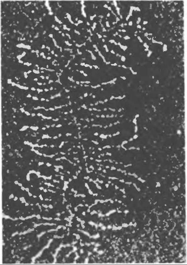
Fig. 54.10. Electron micrograph of a medium-sized proteoglycan aggregate; proteoglycan subunits and the filamentous backbone are particularly well visible. (Reproduced, with permission, from Rosenberg L., Heilman W., Kleinschmidt A. K. Electron microscopic studies of proteoglycan aggregates from bovine articular cartilage. J. Biol. Chem., 1975, 250, 1877.)
Chondroitin sulfates bind tightly to hyaluronic acid with the aid of two link proteins, forming very large aggregates in connective tissue. These aggregates can be observed using an Electron microscope (Fig. 54.10); a schematic representation of them is given in Fig. 54.11.
Link proteins are highly hydrophobic and interact with both hyaluronic acid and the proteoglycan.
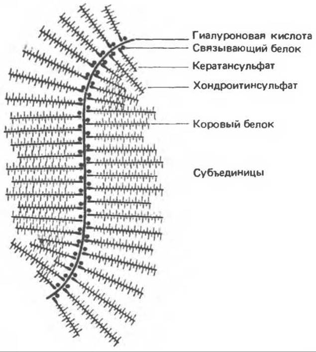
Fig. 54.11. Schematic representation of a proteoglycan aggregate. (Reproduced, with permission, from Lennarz W. J. The Biochemistry of Glycoproteins and Proteoglycans. Plenum Press, 1980.)
Chondroitin sulfates contain 6 types of intersaccharide bonds, and their synthesis therefore proceeds with the participation of 6 different glycosyltransferases—one enzyme for each bond type. In addition, there are 2 types of sulfate esters with sulfate groups at the 4- or 6-positions. These Esterification processes are carried out by two sulfotransferases, whose sulfate-bearing substrate is 3'-phosphoadenosine-5'-phosphosulfate (PAPS).
Keratan Sulfate I and Keratan Sulfate II
As shown in Fig. 54.9, keratan sulfates consist of repeating Gal-GlcNAc disaccharide units and contain sulfate groups at the C-6 position of GlcNAc residues and, occasionally, Gal. The polysaccharide chain in keratan sulfate I is linked to the polypeptide backbone via a GlcNAc-Asn bond. Large amounts of this keratan sulfate are found in the cornea.
Keratan sulfate II is a skeletal proteoglycan found in association with chondroitin sulfate and linked to hyaluronic acid in loose connective tissue. Its polysaccharide chains are attached to the polypeptide backbone through a GalNAc-Thr(Ser) linkage.
Heparin
Heparin is a classic proteoglycan in which several polysaccharide chains are attached to a common protein core. It is found in mast Cell granules and is thus localized intracellularly. Heparin also possesses other Structural and functional properties, some of which are of medical significance. The characteristic Structural Features of heparin are illustrated in Fig. 54.12. The repeating disaccharide unit contains glucosamine (GlcN) and uronic acid. Most of the amino groups of the GlcN residues are present in an N-sulfated form, although a small number of acetylated Amino groups are also present. GlcN also contains a C6-sulfate ester.

Fig. 54.12. Structure of heparin. The polymer segment illustrates typical structural features of heparin, although The sequence of variously substituted repeating disaccharide units is chosen arbitrarily. In addition, non-O-sulfated or 3-O-sulfated glucosamine residues may be present. (Modified, redrawn, and reproduced with permission, from Lindahl U. et al. Structure and Biosynthesis of heparin-like polysaccharides. Fed. Proc., 1977, 36, 19.)
Approximately 90% of the uronic acid is IdUA, with only 10% being GlcUA. Initially, the uronic acid residues are present as GlcUA, but subsequently, after the polysaccharide is formed, a 5-epimerase converts nearly 90% of the GlcUA residues into IdUA residues, which are frequently sulfated at the C-2 position.
The protein moiety of the heparin proteoglycan is unique in that it consists exclusively of Serine and Glycine residues. Approximately two-thirds of the serine residues are linked to polysaccharide chains; their molecular weight typically ranges from 5,000 to 15,000, but can occasionally reach 100,000.
Following polymerization, the polysaccharide chains of heparin proteoglycans undergo a series of modifications:
1) the primary product is unsulfated but fully N-acetylated, yielding the polymer (GlcUA-GlcNAc)n; 2) about 50% of the GlcNAc residues undergo N-deacetylation; 3) the free amino groups of GlcN are sulfated; this is followed by further deacetylation of approximately half of the remaining GlcNAc groups; 4) the N-sulfated polymer serves as a substrate for 5-epimerase, which converts about 90% of the GlcUA residues into IdUA; 5) the newly formed IdUA residues are then subjected to O-sulfation at C-2 positions; 6) the modification is completed by O-sulfation of the GlcN components at C-6 positions.
Heparan sulfate
Heparan sulfate is present on cell surfaces as an extracellular proteoglycan. The polypeptide backbone of heparan sulfate proteoglycan contains an additional amino acid component distinct from that found in heparin. During the modification of its polysaccharide chains, deacetylation of GlcNAc residues occurs to a lesser extent, and therefore it contains fewer N-sulfate groups. Because 5-epimerase (as noted above in the Discussion of heparin modifications) requires N-sulfate substituents as substrates, heparan sulfate differs from heparin by having a lower proportion of IdUA residues and a higher content of GlcUA. Accordingly, the predominant uronic acid in heparan sulfate is GlcUA, whereas in heparin it is IdUA.
Dermatan sulfate
Dermatan sulfate is a widely distributed proteoglycan in animal tissues. Structurally, it shares similarities with both chondroitin sulfate and heparan sulfate. It differs from chondroitin sulfate in that, instead of GlcUA linked to GalNAc via a β-1,3 bond, it contains IdUA linked to GalNAc via an α-1,3 bond. The formation of IdUA, as in the case of heparin and heparan sulfate, occurs via 5-epimerization of GlcUA. Similar to heparin biosynthesis, the epimerization reaction is closely coupled with hexosamine sulfation. Thus, dermatan sulfate contains Two Types of repeating disaccharide units: IdUA–GalNAc and GlcUA–GalNAc.
Last update: 06/08/2026
Editorial and Educational Adaptation: This material has been compiled based on the primary/original source text. The project team performed an editorial review, corrected technical inaccuracies, structured sections, and adapted the content for an educational format.
What was processed:
- elimination of formatting defects (OCR errors, structural breaks, corrupted characters);
- editorial organization of content;
- standardization of terminology in accordance with academic sources;
- verification of factual statements against the original source text.
All mentions of the author, publication year, and origin of the primary text have been preserved in accordance with the source.