Principles of Biochemistry Volume 1 - A. Lehninger 1985
Biomolecules
Enzymes
X-ray diffraction analysis has revealed important structural features of enzymes
X-ray crystallography has provided a wealth of crucial insights into the three-dimensional structures and catalytic mechanisms of Enzymes (Sections 8-2–8-4). It has been applied to a wide range of crystalline enzymes, with these findings complementing and expanding upon data obtained through classical biochemical studies. Some of The most significant breakthroughs achieved via X-ray analysis are explored in Supplement 9-4, which features a veritable "gallery" of enzyme structures.
Supplement 9-4. Structures of Certain Enzymes Determined by X-Ray Diffraction.
A large number of crystalline enzymes have been investigated using X-ray crystallography. The results of these analyses are frequently correlated with data obtained from chemical Methods in 1) determining Amino acid sequences, 2) examining substrate Specificity, 3) studying the action of specific inhibitors, and 4) identifying specific functional groups within the Active Site. To uncover potential links between an enzyme's catalytic activity and its tertiary Structure, representatives from most major enzyme classes have been examined (see Table 9-3). Scaled models of three enzyme molecules are presented here to illustrate key Structural and functional features revealed through the crystallographic analysis of these Proteins.
A. The Lysozyme-Substrate Complex
Although enzyme-substrate complexes are typically transient and rapidly dissociate, it is occasionally possible to synthesize a substrate analog (or employ a chemically modified substrate) that binds to the enzyme's active site and remains stably attached without undergoing catalysis.
Such a substrate was identified for lysozyme, an enzyme that catalyzes the Hydrolysis of specific bonds in bacterial polysaccharide chains. Figure 1 illustrates a scaled representation of the putative normal enzyme-substrate complex for lysozyme. This model was derived from X-ray diffraction data of a crystalline complex formed between lysozyme and a "false" (non-hydrolyzable) substrate analog. These groundbreaking studies were conducted by David C. Phillips and his colleagues at the University of Oxford. The polypeptide backbone, including R-groups and hydrogen atoms, is rendered in red. The segment of the substrate molecule, outlined with black lines, lies within a cleft on the lysozyme surface and is held in place by specific Hydrogen Bonds (depicted as bright red lines) between the enzyme and substrate. The substrate molecule is a polymer consisting of alternating, cyclic units of N-acetylglucosamine and N-acetylmuramic acid linked by glycosidic bonds, denoted A through F (Chapter 11). The specific site where the substrate molecule undergoes Cleavage is indicated by the dashed line.
Class="center">
Fig. 1.
B. The Active Site of Chymotrypsin
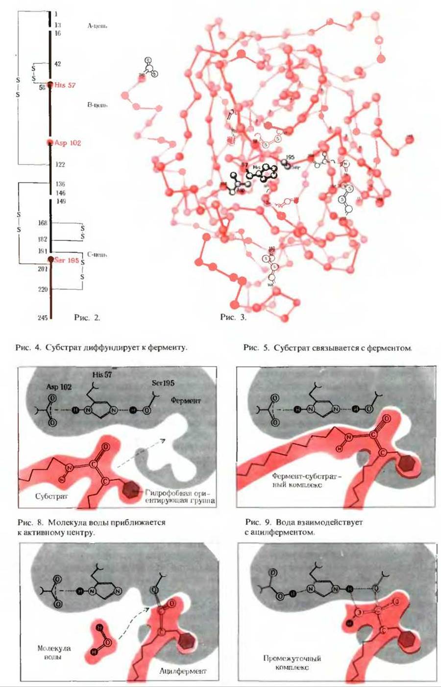
Chymotrypsin is a proteolytic enzyme secreted by the Pancreas into the Small Intestine as an inactive precursor, or zymogen, known as chymotrypsinogen. Chymotrypsinogen consists of a single polypeptide chain of 245 amino acid residues cross-linked by five Disulfide Bonds formed by five cystine residues. It is activated in the small intestine through the action of another protease, Trypsin, which hydrolyzes four peptide bonds and cleaves two dipeptides (at positions 14–15 and 147–148). This yields active chymotrypsin, composed of three polypeptide chains covalently joined by two interchain disulfide bridges—one connecting the A and B chains, and the other linking the B and C chains, as shown in Figure 2. Chymotrypsin activity depends critically on the presence of Histidine 57 and aspartate 102 in the B chain, along with Serine 195 in the C chain. Although these residues are far apart in the linear Amino Acid Sequence (and located on different chains), they are brought into close spatial proximity within the Tertiary Structure of the folded enzyme. This is clearly visible in the space-filling model of the chymotrypsin backbone (Figure 3), which was resolved via X-ray crystallography by David M. Blow and his coworkers at the University of Cambridge. In this illustration, only the R-groups of the three catalytic active-site residues are displayed.
C. Proposed Mechanism of Peptide Hydrolysis by Chymotrypsin
Based on X-ray crystallographic data showing that His-57, Asp-102, and Ser-195 reside in close spatial proximity within the chymotrypsin molecule, a detailed catalytic mechanism involving these residues has been proposed. It is believed that the substrate binds to the active site such that its hydrophobic side chain fits snugly into the nonpolar "pocket" of the enzyme (Figure 4; Section 9-11). Consequently, the target peptide bond is positioned directly adjacent to the hydroxyl group of Ser-195 (Figure 5).
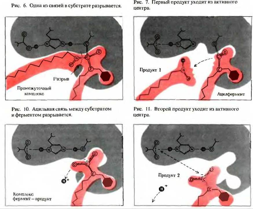
The oxygen atom of this hydroxyl group forms a covalent ester linkage with the carbonyl carbon of the substrate's acyl group, yielding a transient covalent enzyme-substrate intermediate (Figure 6; Section 9-15). The serine hydroxyl readily loses its proton because it is strongly hydrogen-bonded to the electronegative nitrogen of the imidazole ring in His-57. Simultaneously, the peptide bond of the substrate is cleaved, releasing the first reaction product. Following the departure of this product, the acyl group of the substrate remains covalently attached to serine 195, forming an intermediate known as an acyl-enzyme (Figure 7). This ester bond is considerably more labile than the original peptide bond and is readily hydrolyzed to release the second product—the carboxyl portion of the substrate. During this step, a proton is transferred back to the serine residue (Figures 8 and 9), generating an enzyme-product complex (Figure 10). The second product then dissociates from the active site, completing the catalytic cycle (Figure 11). The acyl-enzyme intermediate represents a cornerstone of this Covalent Catalysis pathway, while the imidazole group of histidine 57 acts as a general acid-base catalyst facilitating proton transfer.
It has been suggested that the primary role of the negatively charged aspartate 102 residue is to enhance the basicity and mobility of the histidine 57 imidazole ring, thereby optimizing its ability to Abstract the proton from serine 195. However, some debate persists as to whether such a formal "charge Relay system" truly operates, given the relatively distance between Asp-102 and His-57. Regardless of the exact electrostatic role of aspartate 102, experimental evidence confirms that it is strictly essential for catalytic activity.
D. Induced Fit Between Hexokinase and D-Glucose
Hexokinase catalyzes the phosphorylation of D-glucose and other hexoses by ATP:
ATP + D-glucose -> ADP + D-glucose-6-phosphate,
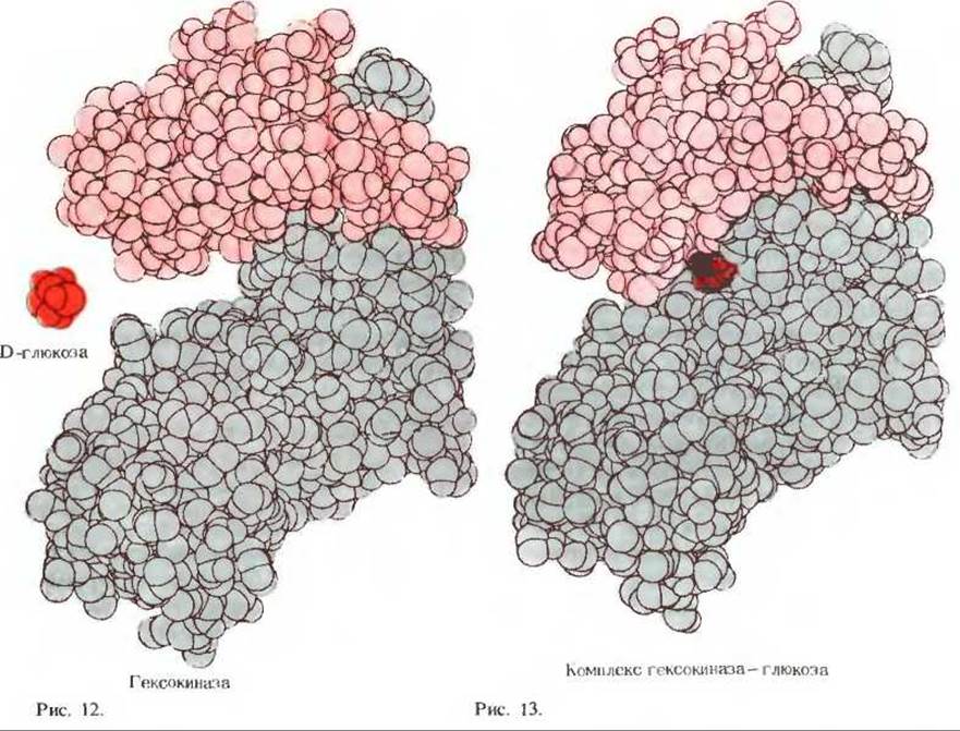
consisting of two polypeptide subunits, as illustrated by the space-filling molecular model in Figure 12. A free molecule of D-glucose (colored dark red) is shown alongside the "unliganded" hexokinase molecule. When D-glucose binds to the active site in the absence of the second substrate, ATP, the two subunits undergo a dramatic conformational shift, closing around the glucose molecule to enclose it securely within the active-site pocket (Figure 13). This transition involves a substantial reorganization of the enzyme's quaternary structure, as hexokinase undergoes an induced fit upon binding the substrate alone. This stable binary complex has been successfully crystallized, and its X-ray structure was elucidated by Thomas A. Steitz at Yale University. When both ATP and glucose are present simultaneously, they bind to their respective sites, initiating the catalytic transfer to yield ADP and glucose-6-phosphate, which then dissociate. Following product release, the enzyme relaxes back to its initial open conformation, ready to embark on a new catalytic cycle.
First and foremost, X-ray crystallography enables the precise Determination of the secondary, tertiary, and quaternary structures of various enzymes, allowing direct comparisons with non-catalytic Globular proteins. Such comparative analyses have revealed no unique three-dimensional architectural motifs that fundamentally distinguish enzymes from non-enzymatic proteins. Nonetheless, enzymes belonging to the same functional family—such as Kinases that catalyze phosphoryl transfer from ATP to specific acceptors—often share conserved structural features essential for their function.
Furthermore, crystallographic studies have successfully mapped the active sites of numerous enzymes. The active site frequently manifests as a cleft or depression on the enzyme's surface whose geometry is precisely complementary to that of the incoming substrate. In some enzymes, active sites are lined by loops of polypeptide chains adopting a beta-conformation, whereas in others they form hydrophobic pockets rich in charged polar amino acid residues. In favorable cases, X-ray analysis has successfully captured high-resolution snapshots of enzyme-substrate complexes, a prime example being lysozyme (Supplement 9-4, A), which cleaves specific bonds within the polysaccharide backbone of bacterial Cell walls.
The synergy of X-ray crystallography and chemical modification studies has illuminated the intricate Topography of the chymotrypsin active site, which comprises three polypeptide chains held together by interchain disulfide bonds between cystine residues (Supplement 9-4, B). Chemical labeling experiments demonstrated that the irreversible inactivation of chymotrypsin by diisopropylfluorophosphate results from the Covalent Modification of a specific serine residue at position 195, thereby confirming its role as a catalytic active-site residue. Additional biochemical studies established that histidine 57 and aspartate 102 also participate directly in catalysis. Although these residues are distant from one another in the primary sequence and reside on separate polypeptide chains, X-ray diffraction data revealed that in the folded, native tertiary structure of chymotrypsin (Supplement 9-4, B), they are brought into remarkably close spatial alignment. These precise structural insights, combined with classical biochemical data, have led to robust models for chymotrypsin catalysis (Supplement 9-4, B) while ruling out alternative mechanisms incompatible with the active-site geometry. Although certain minute details of chymotrypsin action remain under investigation, its catalytic mechanism is currently understood in greater depth than that of almost any other enzyme.
X-ray crystallographic investigations have unveiled another fundamental paradigm in enzymology: the induction of significant conformational changes within the enzyme molecule upon substrate binding and subsequent catalysis. A striking illustration of this phenomenon is hexokinase (Section 9-3), which catalyzes the phosphorylation of D-glucose by ATP. As detailed in Supplement 9-4, D, the binding of the relatively small glucose molecule to the hexokinase active site triggers a massive closure of the two polypeptide subunits, which clamp down around the sugar like a pair of jaws to position it optimally for nucleophilic attack by ATP. It is widely thought that this induced conformational adjustment strains the bound glucose molecule, pushing its geometry closer to that of the Transition State.
Last update: 06/08/2026
Editorial and Educational Adaptation: This material has been compiled based on the primary/original source text. The project team performed an editorial review, corrected technical inaccuracies, structured sections, and adapted the content for an educational format.
What was processed:
- elimination of formatting defects (OCR errors, structural breaks, corrupted characters);
- editorial organization of content;
- standardization of terminology in accordance with academic sources;
- verification of factual statements against the original source text.
All mentions of the author, publication year, and origin of the primary text have been preserved in accordance with the source.