Principles of Biochemistry, Volume 1 - A. Lehninger 1985
Biomolecules
Globular Proteins: Structure and Function of Hemoglobin
Globular proteins of different types have distinct structures
Are all other Globular Proteins folded in the same way as Myoglobin? This question has already been answered, as X-Ray Diffraction Analysis has established the Tertiary Structure of A number of other small, single-chain proteins. Particularly interesting are the results obtained from studying the mitochondrial protein cytochrome c, which serves as an electron carrier. The Amino Acid Sequence of cytochrome c has been determined for more than 60 species (Section 6.10). Like myoglobin, cytochrome c is a small heme-containing protein (mol. wt. 12,400) with a single polypeptide chain of approximately 100 amino acid residues and a single heme group, which in this case is covalently linked to the polypeptide. Like myoglobin, cytochrome c is folded into a compact globule, with most of its hydrophilic R-groups located on the outside and most of its hydrophobic R-groups on the inside of the globular structure. Since both cytochrome c and myoglobin are heme proteins, one might expect them to have similar tertiary structures. However, this is not the case. X-ray diffraction analysis of cytochrome c has shown that it has a completely different three-dimensional structure (Fig. 8-5 and Table 8-2). While nearly 80% of The amino acid residues in myoglobin are in α-helical segments, α-helices account for only 40% of the residues in cytochrome c. The remainder of the cytochrome c polypeptide chain contains various bends, turns, irregular coils, and segments in an extended conformation. Thus, although both cytochrome c and myoglobin are heme-containing proteins, they differ greatly in both secondary and tertiary structures, as well as in their Amino acid sequences, in keeping with their completely different biological Functions.
Class="center">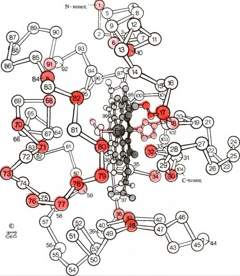
Fig. 8-5. Backbone of the cytochrome c molecule. The covalently bound heme group, shown in gray, is located in a cavity inside the molecule. Invariant amino acid residues are highlighted in red.
In cytochrome c, the fifth and sixth coordination positions of the iron atom are occupied by the R-groups of residues 18 and 80. In normally functioning cytochrome c participating in the Electron Transport Chain, the iron atom alternates between the ferrous and ferric states, i.e., the [Fe(II)] and [Fe(III)] forms.
For comparison, we can examine the tertiary structure of two other small proteins. Lysozyme is an enzyme found in egg white as well as in human tears. It catalyzes the hydrolytic Cleavage of complex Polysaccharides present in The Cell walls of certain Bacteria. Lysozyme is so named because it causes lysis, or dissolution, of bacterial cell walls, and thus can act as a bactericidal agent. Like myoglobin and cytochrome c, lysozyme has a compactly folded conformation, with most of its hydrophobic R-groups located inside the globular structure, where they are shielded from contact with Water, while the hydrophilic groups project outward into the aqueous environment. About 40% of the 129 amino acid residues of lysozyme are in α-helical segments (Table 8-2), which line a long cleft on one side of the molecule. This cleft represents the Active Site of the enzyme. As we will see in Chapter 9, every enzyme has an active site that binds the substrate—the molecule acted upon by the enzyme. During catalysis, the bacterial polysaccharide substrate of lysozyme fits snugly into this cleft. Figure 8-6 shows the space-filling model of lysozyme, illustrating the compact Introduction/12.html">Structure of Globular proteins.
Table 8-2 Approximate content of α-helices and β-structure in some single-chain proteins1)
|
Protein |
Total number of residues |
Residues, % |
|
|
α-helices |
β-structure |
||
|
Myoglobin |
153 |
78 |
0 |
|
Cytochrome c |
104 |
39 |
0 |
|
Lysozyme |
129 |
40 |
12 |
|
124 |
26 |
35 |
|
|
241 |
14 |
45 |
|
|
Carboxypeptidase |
307 |
38 |
17 |
1) Portions of polypeptide chains that are neither α-helices nor β-structures consist of bends or reverse turns, as well as irregularly coiled or extended segments. The α-helical and β-conformation segments sometimes have slightly distorted dimensions and geometry, differing from the corresponding standard parameters. Data from Cantor, C. R. and Schimmel, P. R., Biophysical Chemistry, pt. 1, p. 100, Freeman, San Francisco, 1980.

Fig. 8-6. Space-filling model of a lysozyme molecule tightly binding a polysaccharide substrate molecule (shown in red). Note the extremely compact shape of the lysozyme molecule, leaving almost no empty space inside. For another representation of the lysozyme molecule, see Fig. 1 in Box 9-4 in Chapter 9.
Ribonuclease, another small globular protein, is an enzyme secreted by Pancreas Cells into the Small Intestine, where it catalyzes the Hydrolysis of certain bonds in ribonucleic acid molecules present in digested food. The tertiary structure of ribonuclease, determined by X-ray diffraction analysis (Fig. 8-7), is characterized by having very few α-helical regions in its polypeptide chain, but rather a relatively large number of segments in the β-conformation. In this respect, ribonuclease differs from myoglobin, cytochrome c, and lysozyme. However, like lysozyme, it contains four cystine residues that form covalent Disulfide Bonds between loops of the polypeptide chain. This stabilizes the entire native tertiary STRUCTURE OF THE enzyme (Fig. 8-7). Such intrachain disulfide bonds are found in many proteins, especially those that function extracellularly.

Fig. 8-7. Conformation of the ribonuclease molecule determined by X-ray diffraction analysis. Dashed lines indicate Hydrogen Bonds between loops of the polypeptide chain arranged as a β-pleated sheet. The cleft in the middle of the upper part of the molecule serves as the substrate-binding site. The arrangement of intrachain disulfide cross-links is shown in Fig. 8-8.
It is quite clear that these four small, single-chain globular proteins differ significantly from one another (see Table 8-2). They contain different numbers of α-helical regions and segments in the β-conformation; consequently, they fold differently in space. These proteins also differ in their amino acid sequences and perform completely different biological functions. From data obtained through X-ray diffraction studies and amino acid sequencing of many types of globular proteins, it is now well established that each type of protein has a characteristic three-dimensional conformation specifically tailored to perform its particular biological function.
Last update: 06/08/2026
Editorial and Educational Adaptation: This material has been compiled based on the primary/original source text. The project team performed an editorial review, corrected technical inaccuracies, structured sections, and adapted the content for an educational format.
What was processed:
- elimination of formatting defects (OCR errors, structural breaks, corrupted characters);
- editorial organization of content;
- standardization of terminology in accordance with academic sources;
- verification of factual statements against the original source text.
All mentions of the author, publication year, and origin of the primary text have been preserved in accordance with the source.