LEHNINGER PRINCIPLES OF BIOCHEMISTRY - VOL. 2. BIOENERGETICS AND METABOLISM - 2014
PART II. BIOENERGETICS AND METABOLISM
19. OXIDATIVE PHOSPHORYLATION AND PHOTOPHOSPHORYLATION
19.10. The Evolution of Oxygenic Photosynthesis
The Emergence of oxygenic Photosynthesis on Earth 2.5 billion years ago was a pivotal event in the Evolution of the biosphere. Before this time, the Earth's atmosphere and oceans were virtually devoid of molecular oxygen and the ozone layer that protects living organisms from solar ultraviolet radiation. Oxygenic photosynthesis provides a virtually inexhaustible supply of reducing equivalents to drive The formation of Organic compounds via reductive biosynthetic pathways. The corresponding reaction mechanisms allow organisms to use O2 as a terminal electron acceptor in high-energy electron-transfer reactions driven by organic substrates, and to harness the energy of oxidation to sustain their own METABOLISM. The complex photosynthetic apparatus of modern vascular plants is a supreme evolutionary achievement; later, relationships were established between Eukaryotic Cells and cyanobacteria acting as endosymbionts.
Chloroplasts Arose from the Endosymbiotic Evolution of Bacteria
The chloroplasts of modern organisms share several features with Cell/35.html">Mitochondria; they are believed to have arisen through the same mechanism that produced mitochondria, namely, endosymbiosis. Like mitochondria, chloroplasts contain their own set of genes in DNA molecules and a protein-synthesizing machinery. Some chloroplast Proteins are encoded by chloroplast genes and synthesized within the chloroplasts, whereas others are encoded by nuclear DNA, synthesized outside the chloroplasts, and subsequently imported into them. These topics are discussed in detail in Chapter 27 (Vol. 3). Plant Cell Growth and Division trigger chloroplast division, during which DNA Replication occurs and is partitioned between the daughter chloroplasts. The molecular machinery and mechanism of light-energy harvesting, the electron-transfer chain, and ATP Synthesis in photosynthetic bacteria and chloroplasts share striking similarities. Based on these findings, the widely accepted hypothesis of the endosymbiotic origin of chloroplasts posits that modern plant cells are descendants of photosynthetic bacteria that once invaded eukaryotic cells and became stably established within them during evolution (see Fig. 1-36, Vol. 1).
At least half of all photosynthesis on Earth is carried out by microorganisms—Algae, lower eukaryotes, and photosynthetic bacteria. In cyanobacteria, the oxygen-evolving system resembles that of green plants, consisting of coordinately functioning Photosystems II and I, one of which includes a Water-splitting complex. All other photosynthetic bacteria possess a single photosystem; they do not split water and do not produce oxygen. Furthermore, many photosynthetic bacteria are obligate anaerobes—meaning they cannot tolerate oxygen at all—and use Inorganic Compounds rather than water as electron (hydrogen) Donors. For example, green sulfur bacteria use hydrogen sulfide as their hydrogen donor:
Class="center">
Instead of molecular oxygen, these bacteria release elemental sulfur, which is the oxidation product of H2S. The sulfur is subsequently oxidized to SO2-4. Other photosynthetic bacteria use organic compounds, such as lactate, as electron (hydrogen) donors:

Accumulated empirical evidence indicates that the fundamental processes of photosynthesis in plants and bacteria are largely identical, despite utilizing different hydrogen donors. This similarity becomes apparent when the overall equation for photosynthesis is written in a more general form:

where H2D is the hydrogen donor and D is its oxidized form. Depending on the species of photosynthetic Organism, H2D may be water, hydrogen sulfide, lactate, or other organic compounds. Most likely, the earliest photosynthetic organisms on Earth were bacteria that utilized H2S as an electron donor.
The ancient relatives of modern cyanobacteria may have originated from a combination of genetic material from two surviving types of photosynthetic bacteria: purple bacteria (featuring electron transport analogous to PSII) and green sulfur bacteria (featuring electron transport analogous to PSI). Bacteria with two independent photosystems could use one under certain environmental conditions and the other under different ones. Over time, a mechanism evolved that linked the Two Photosystems, allowing both to function simultaneously; a system akin to PSII in modern cyanobacteria acquired the capacity to split water molecules.
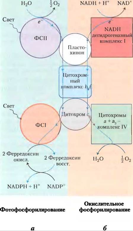
Modern cyanobacteria can synthesize ATP via Oxidative Phosphorylation or Photophosphorylation, even though they lack Mitochondria and chloroplasts. The enzymatic machinery required for both processes is localized in a highly invaginated Plasma Membrane (see Fig. 1-6, Vol. 1). Three protein components function in both pathways, pointing to a common evolutionary origin (Fig. 19-65). First, the cytochrome b6f complex pumps protons from plastoquinone to cytochrome c6 during photosynthesis, and from ubiquinone to cytochrome c6 during oxidative phosphorylation (in mitochondria, this role is performed by cytochrome bc1). Second, cytochrome c6, which is homologous to mitochondrial cytochrome c, transfers electrons from complex III to complex IV in cyanobacteria; it can also shuttle electrons from the cytochrome b6f complex to PSI (in plants, this role is played by plastocyanin). Thus, there is functional Homology between the cytochrome b6f complex of cyanobacteria and the mitochondrial cytochrome bc1 complex, as well as between cyanobacterial cytochrome c6 and plant plastocyanin. The third conserved component is ATP synthase, which participates in both OXIDATIVE PHOSPHORYLATION AND photophosphorylation in cyanobacteria and is also present in the mitochondria and chloroplasts of photosynthetic eukaryotes. The Structure and remarkable MECHANISM OF ACTION of this enzyme have been strictly conserved throughout evolution.
Figure 19-65. The dual role of cytochrome b6f and cytochrome c6 in cyanobacteria reflects their evolutionary origins. Cyanobacteria utilize the cytochrome b6f complex, cytochrome c6, and plastoquinone for both oxidative phosphorylation and photophosphorylation. (a) During photophosphorylation, electrons flow (from top to bottom) from water to NADP+. (b) During oxidative phosphorylation, electrons flow from NADPH to O2. Both processes are coupled to proton translocation across the membrane via Q-cycle reactions.
In Halophilic Bacteria, a Single Protein Absorbs Sunlight and Pumps Protons Out of The Cell, Generating the Driving Force for ATP Synthesis
Some modern archaea employ a somewhat different mechanism for converting light energy into an electrochemical gradient. The ancient evolutionarily conserved halophilic (salt-loving) bacterium Halobacterium salinarum harnesses solar energy in a manner entirely distinct from true photosynthetic organisms. This peculiar bacterium thrives exclusively in saline water bodies (such as salt ponds and hypersaline lakes, e.g., the Great Salt Lake and the Dead Sea), where evaporation can drive salt concentrations above 4 M. These bacteria are incapable of surviving at NaCl concentrations below 3 M. As aerobes, they typically rely on oxygen to oxidize their organic "fuel." However, the solubility of oxygen in hypersaline waters is extremely low. Consequently, halophilic bacteria must occasionally resort to an alternative energy source: sunlight.
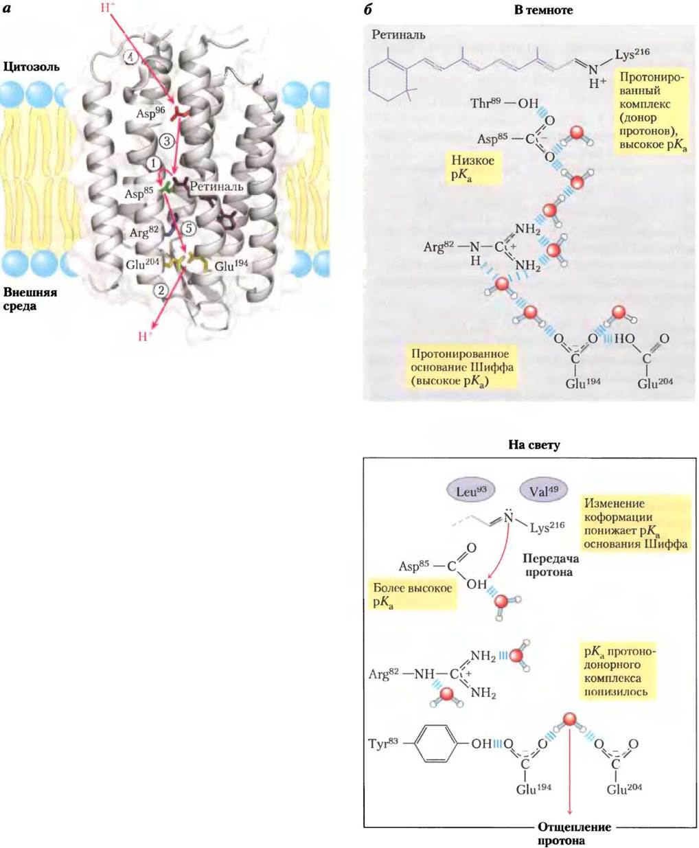
The cytoplasmic membrane of H. salinarum contains patches of light-absorbing pigments composed of the protein Bacteriorhodopsin, which contains a retinal residue—a vitamin A aldehyde—as a prosthetic group (see Fig. 10-21, Vol. 1). Upon illumination, the all-trans-retinal bound to bacteriorhodopsin absorbs a photon and undergoes photoisomerization to 13-cis-retinal. The thermal relaxation back to the original all-trans configuration releases energy, which is coupled to the pumping of protons from the cell into the extracellular medium. The bacteriorhodopsin molecule consists of a mere 247 amino acid residues and Functions as a primitive light-driven protein proton pump. Figure 19-66a depicts the three-dimensional structure of bacteriorhodopsin in the dark and in the light, illustrating The pathway of proton translocation across the membrane via a sequence of coordinated "hops." In bacteriorhodopsin, the retinal chromophore is attached to the ε-amino group of a Lys residue via a Schiff base. In the dark, the nitrogen atom of the Schiff base is protonated. Upon illumination, photoisomerization of retinal lowers the pKa of the Schiff base, causing a proton to dissociate from the nitrogen atom and transfer to an adjacent Asp residue. A series of proton "hops" ensues—resulting in The transfer of a proton from the inner membrane surface to the extracellular medium, thereby generating a transmembrane pH gradient with a more acidic environment outside (Fig. 19-66c).
Figure 19-66. Evolution of a second light-driven proton-pumping mechanism in halophilic archaea. (a) Bacteriorhodopsin (Mr = 26,000) from Halobacterium halobium contains seven transmembrane α-helices (PDB ID 1C8R). The all-trans-retinal chromophore (colored purple) is linked to the ε-amino group of a Lys residue, forming a Schiff base within the membrane. Asp and Glu residues located within the α-helices, along with several coordinated water molecules, facilitate proton hopping during transmembrane transport (indicated by red arrows). Steps (1) through (5) outline the proton translocation cycle discussed in the text. (b) In the dark (top right), the Schiff base linking the bacteriorhodopsin chromophore to the Lys residue is protonated. In the light (bottom right), photoisomerization of retinal induces subtle protein conformational changes—altering distances between the Schiff base and neighboring amino acid residues—which lowers the pKa of the Schiff base and triggers proton release to the carbonyl group of the adjacent Asp85 residue. This process (step (1) in Fig. a) induces proton hopping through water molecules coordinated within the protein interior, ultimately resulting (stage (2)) in proton release from a pair of amino acid residues, Glu194-Glu204, on the outer surface of the cell. Next (stage (3)), the Schiff base reprotonates by acquiring a proton from Asp96, which in turn captures a proton from the Cytosol (stage (4)). Finally, Asp85 transfers a proton to the Glu204-Glu194 amino acid pair (stage (5)), resetting the system for a new cycle of transmembrane proton pumping into the extracellular medium.
Because the extracellular H+ concentration becomes higher than the intracellular concentration, these ions tend to diffuse back into the cell through membrane-bound ATP synthase enzyme complexes resembling those found in mitochondria and chloroplasts. As H+ ions flow through this bacterial ATPase, they release energy that is harnessed to synthesize ATP from ADP and inorganic phosphate. In this manner, in the absence of O2, halophilic bacteria capture light energy in the form of ATP to Supplement the ATP generated via oxidative phosphorylation when oxygen is available. However, halophilic bacteria do not evolve oxygen nor do they perform photoreduction of NADP+. Light-dependent energy Transduction in halophilic bacteria is simpler than in cyanobacteria or plants. Understanding the Molecular structure and mechanism of action of bacteriorhodopsin—the simplest known light-driven proton pump—promises to provide deeper insights into the operation of more complex H+-pumping systems. Bacteriorhodopsin
Summary of Section 19.10 The Evolution of Oxygenic Photosynthesis
■ Modern cyanobacteria evolved from an ancient organism possessing two photosystems: a system of one type can be found today in purple bacteria, and a System of the other type in green sulfur bacteria.
■ Many photosynthetic organisms derive electrons for photosynthesis not from water, but from other donors such as H2S.
■ Cyanobacteria featuring two coupled photosystems and The ability to split water molecules with the release of oxygen into the atmosphere appeared on Earth approximately 2.5 billion years ago.
■ Chloroplasts, much like mitochondria, originated through the evolutionary process of endosymbiosis with bacteria that once invaded the cells of ancient eukaryotes. Consequently, bacteria, cyanobacteria, mitochondria, and chloroplasts share a common ancestor of the ATP synthase enzymatic complex and, therefore, a common catalytic mechanism.
■ A completely different mechanism for converting light energy into a proton gradient emerged in modern archaea, in which retinal serves as the light-absorbing pigment.
Last update: 06/08/2026
Editorial and Educational Adaptation: This material has been compiled based on the primary/original source text. The project team performed an editorial review, corrected technical inaccuracies, structured sections, and adapted the content for an educational format.
What was processed:
- elimination of formatting defects (OCR errors, structural breaks, corrupted characters);
- editorial organization of content;
- standardization of terminology in accordance with academic sources;
- verification of factual statements against the original source text.
All mentions of the author, publication year, and origin of the primary text have been preserved in accordance with the source.