Chemistry and Biology of Proteins - F. Haurowitz 1953
Conjugated Proteins
Lipoproteins
Proteins can form both soluble and insoluble complexes with Lipids. The first type includes Lipoproteins found in Blood and other animal Body Fluids. Blood Plasma, despite being a transparent fluid, contains 0.5–0.7% of insoluble lipids. A significant portion of these lipids cannot be extracted from plasma using ether or other non-polar Solvents traditionally employed for this purpose. Machebœuf views this as evidence that the specified fraction of lipids is bound to proteins, forming complexes that he termed “synapses” [3]. These lipoprotein complexes are precipitated by conventional salting-out with ammonium sulfate [4]. Certain amounts of lipids can also be detected in protein fractions obtained electrophoretically [5]. Lipoprotein complexes are dissociated at room Temperature by ethyl alcohol and acetone; furthermore, the majority of lipids detached from the complex following alcohol Treatment can be extracted with ether. To prevent Protein Denaturation, it is recommended to carry out the Cleavage of the lipoprotein complex with alcohol and ether at low temperatures [6] or by repeatedly freezing and thawing these complexes in the presence of ether [7].
If blood serum is extracted with ether at —70°, all the Cholesterol passes into the extract, whereas Phospholipids remain bound to proteins [8]. Similar results can be achieved by using cationic detergents [9] (see Chapter X).
Fractionation of human blood serum with ethyl alcohol (see Chapter VIII) yields primarily two lipoproteins: ß1-lipoprotein from fraction III—0 and a1-lipoprotein from fraction IV—1, accounting for 5% and 3% of the total Plasma Proteins, respectively [10—12]. ß1-Lipoprotein constitutes about 70% of all plasma lipids. It contains 25% protein, 30% phospholipids, and 45% cholesterol and cholesterol esters. Its molecular weight is 1,300,000. a1-Lipoprotein contains 65% protein and 35% lipids [10—12]. Its molecular weight is 200,000.
The bonds linking proteins to lipids within lipoprotein complexes presumably belong to various types. To date, preparing such complexes simply by mixing proteins with lipids has been unsuccessful. These complexes likely form in the Organism at the moment the protein macromolecule is synthesized. One may conceive that lipids penetrate between the folding peptide chains during this process. Consequently, the dissociation of such a complex can occur only after the unwinding of the peptide chains. Another possibility to consider is that lipids form the core of the micelle and are enveloped by proteins [13].
Until recently, blood phospholipids were thought to consist mainly of lecithins and cephalins. Lately, however, it has been established that blood phospholipids also incorporate sphingomyelin [14, 15]. The Nature of the bond between phospholipids and proteins likewise remains not fully determined. Cephalins, which contain phosphoric acid and Serine residues, exhibit acidic properties and are capable of forming insoluble compounds with basic protamines and egg albumin [16].
Class="center">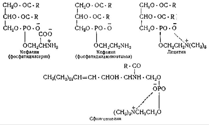
The ratio of salmine to cephalin in the insoluble salmine-cephalin complex is approximately 3:1. Lecithin and sphingomyelin are less acidic compounds than cephalins, since the phosphoric acid in them is partially neutralized by the ammonium group of Choline. Consequently, neither protamines nor other proteins are precipitated by lecithin. This, however, does not imply that lecithin and sphingomyelin cannot form electrovalent bonds with proteins. Such bonds may arise between the phosphoric acid anion and the cationic groups of proteins, or between the quaternary ammonium group of choline and the acidic groups of proteins.

In addition to these electrovalent bonds (salt bridges), one can envision The formation of bonds involving both polar and non-polar groups of the proteins and lipids making up the complex. At present, however, nothing definitive can be stated regarding these bonds in soluble blood plasma lipoproteins.
A distinct group comprises lipoproteins that form part of Cell membranes and cytoplasmic structural elements (Mitochondria and microsomes). Although the mere presence of lipids in these cellular elements has been known for a long time, we still know almost nothing about the nature of their bonding. Unlike the lipids of adipose tissue, the lipids composing cell membranes and cytoplasmic structural elements are stained by neither Sudan III, scarlet red, nor Nile blue. Therefore, they are often referred to as “masked lipids.” The inability of these lipids to combine with lipophilic Dyes serves as strong evidence that they are bound to proteins. Liver cell mitochondria contain 15—20% lipids [17]. Submicroscopic particles (microsomes) isolated from the liver or Pancreas by high-speed centrifugation contain up to 40—50% lipids bound to Nucleoproteins [18] (see also p. 260).
Certain lipoproteins are BIOLOGICALLY ACTIVE SUBSTANCES. For instance, lipoproteins from animal Tissues exhibit thromboplastic properties and play a major role in Blood Coagulation (see Chapter VIII). The endotoxins of Shigella and other Bacteria are likewise lipoproteins (see Chapter XV).
Proteins can also form compounds with carotenes1. The coloration of crustaceans is due to cyanin [19], a lipoprotein containing the carotene astaxanthin (5,5'-dioxy-4,4'-diketo-ß-carotene) [20]. This lipoprotein is extracted from lobster shells with citric acid [19]. A similar lipoprotein, ovoverdine, forms the green pigment of lobster eggs. The Molecular Weight of this lipoprotein, which contains one molecule of astaxanthin per protein molecule, is approximately 300,000. Its isoelectric point lies at pH 6.7 [21]. As long as the carotene incorporated into this lipoprotein remains bound to the protein, it is protected against oxidation; upon heating, however, the protein is denatured and the carotene is released as a red pigment. Carotene-containing lipoproteins have also been found in green grasshoppers [22] and presumably occur in other insects as well.
1 Regarding the binding of blood serum β-globulins to carotene and vitamin A, see the footnote on p. 177. — Ed. note.
Among the carotene-containing lipoproteins, of greatest interest is the lipoprotein constituting the rods of the retina—visual purple [23, 24]. This lipoprotein, absent in cones, is known to influence night visual acuity (Kühne, 1879). Visual purple can be extracted from the retina by compounds containing hydrophilic and lipophilic groups, such as Bile, digitonin [25], and invert soaps [26]. Stable solutions of visual purple can be obtained by extraction with a 75% aqueous glycerol solution [27]. Solutions of visual purple must be prepared in the dark, as they are instantly bleached upon exposure to light (whereby the pink color of the solution first turns yellow, followed by complete bleaching). According to Wald [28], the intermediate yellow color is due to the presence of the pigment retinene. This pigment, which is an aldehyde, can be obtained from vitamin A by its oxidation with manganese dioxide [29]. The molecular weight of dissolved visual purple, determined by the sedimentation method, is 240,000 [30]; whereas determinations by the diffusion method yielded values on the order of 600,000—800,000 [31]. The isoelectric point of this protein lies at pH 4.47—4.57 [27].
Animal visual purple incorporates two different carotenoids. The visual purple of chickens, frogs, and marine fish contains rhodopsin, which upon illumination converts into retinene—a vitamin A derivative. The retina of freshwater fish contains a purple-colored porphyropsin, which upon illumination transforms into retinene2—a vitamin A2 derivative [24]. The absorption maxima of rhodopsin and porphyropsin lie at 500 and 522 mµ, respectively [24].
Rods contain 5—10% visual purple, which roughly corresponds to a content of 109 molecules of this protein per single rod [27].
The Mechanism of the reactions occurring upon illumination is not yet fully elucidated. According to Wald [24, 28], exposure to light causes The breakdown of visual purple into protein and retinene. The Regeneration of Visual purple is an aerobic process, with oxygen consumption by the retina being higher in the dark than in the light [32]. Several authors [26, 33] believe that the photoreaction is accompanied by protein denaturation. This is supported by the fact that dry visual purple exhibits the highest stability. The red residue obtained by drying visual purple in a vacuum over calcium chloride is sufficiently stable toward daylight [34].
The primary product formed upon illumination of the carotenoid component of visual purple has been termed transient orange. It is stable at temperatures around 0°, whereas at room temperature it converts into indicator yellow, which loses its color in alkaline solution [35]. Indicator yellow is a conjugated protein in which the aldehyde group of retinene is likely linked to an amino group of the protein moiety [36]. Photolysis of dry visual purple does not yield vitamin A [34, 37]. Treatment with gasoline or chloroform does not alter visual purple, whereas treatment with ethyl or methyl alcohol instantly transforms it into a yellow substance. Since retinene is yellow and vitamin A is colorless, neither the former nor the latter can be regarded as the prosthetic group of visual purple; thus, we are still unable to say anything about The Structure of the substance responsible for the intense color and photosensitivity of visual purple.
The coloration of carotenes is primarily due to their chain length and the number of double bonds within that chain. It is highly probable that two or more retinene molecules combine with each other, forming a red-colored carotene derivative that is incorporated into visual purple [28, 36]. Unlike rods, retinal cones contain pigments responsible for daylight Vision. These pigments are also carotenoids, possibly bound to proteins [19].
Last update: 06/08/2026
Editorial and Educational Adaptation: This material has been compiled based on the primary/original source text. The project team performed an editorial review, corrected technical inaccuracies, structured sections, and adapted the content for an educational format.
What was processed:
- elimination of formatting defects (OCR errors, structural breaks, corrupted characters);
- editorial organization of content;
- standardization of terminology in accordance with academic sources;
- verification of factual statements against the original source text.
All mentions of the author, publication year, and origin of the primary text have been preserved in accordance with the source.