Biochemistry and Molecular Biology - Belyasova N.A. 2002
Molecular Foundations and Mechanisms of Heredity
Maintenance of Stability and Variability of Genomes
DNA Repair
Despite the high fidelity of DNA-replicating Enzymes and the existence of proofreading mechanisms, errors still occur during the synthesis of new DNA strands due to the incorporation of non-complementary NUCLEOTIDES. Furthermore, cellular DNA molecules are exposed to various Physical and Chemical factors that disrupt their Structure. The most frequent types of DNA damage include the following:
— Cleavage of the (β-N)-glycosidic bonds between Purines and deoxyribose (depurination), which is most often a consequence of elevated Temperature. In a human Cell, between 5,000 and 10,000 depurination events occur per day;
— Spontaneous deamination of cytosine and adenine residues, yielding uracil and hypoxanthine residues, respectively (approximately 100 events per genome per day);
— alkylation of nitrogenous bases by a special Class of chemical compounds (alkylating agents);
— intercalation (insertion) of certain compounds between adjacent Base Pairs;
— formation of covalent cross-links between DNA strands induced by bifunctional agents;
— formation of cyclobutane dimers (Fig. 2.2) between adjacent Pyrimidines in the strand upon absorption of ultraviolet (UV) light.
Most of these lesions interfere with METABOLISM/36.html">DNA Replication and Gene Expression; for instance, each thymine dimer in E. coli DNA delays replication by 10 seconds. Moreover, such lesions serve as a source of Mutations if they are not repaired prior to the onset of DNA replication.
Typically, such damage occurs in only one of the DNA strands, while the opposite strand in most cases retains the "correct" sequence, which can serve as a template for error correction. Thus, The Double Helix of DNA, along with the encoded information regarding The structure of repair enzymes, enables a unique error-correction mechanism known as DNA Repair, which is characteristic exclusively of this class of molecules.
Organisms possess A wide variety of repair systems and mechanisms, some of which are specific to a single type of lesion, while others are less specific. For convenience, all currently known repair processes can be divided into two categories: 1) those that do not require replication and represent direct correction of DNA Lesions; 2) more complex processes involving repair replication. Repair mechanisms have been studied most thoroughly in relation to the correction of UV-induced damage, namely pyrimidine dimers (Fig. 2.2).
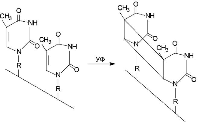
Fig. 2.2. The formation of thymine dimers in one of the DNA strands involves the creation of a cyclobutane ring (consisting of four carbon atoms) through the interaction of atoms from adjacent pyrimidine bases.
Since the most well-known processes repairing UV-induced damage involve UV-dependent enzymes, repair mechanisms are also classified into light repair (which can occur only in the presence of visible light) and dark repair (which does not require visible light).
Mechanisms of direct damage reversal include the dealkylation of guanine residues and the monomerization of cyclobutane dimers between adjacent pyrimidine bases. The dealkylation of methylguanine residues is a form of dark repair and is mediated by enzymes present in bacterial and mammalian Cells. O6-methylguanine-DNA alkyltransferase catalyzes The transfer of alkyl groups to The sulfhydryl groups of Cysteine residues within the enzyme (Fig. 2.3).
The cleavage of dimers between pyrimidine nucleotides occurs during photoreactivation—the restoration of UV-damaged DNA molecules through subsequent exposure to visible light (light repair). Non-enzymatic short-wavelength photoreactivation is known, which involves the monomerization of dimers under ultraviolet radiation at a wavelength of 240 nm, as well as enzymatic photoreactivation. The latter is generally what is meant by photoreactivation proper. This process requires visible light with a wavelength of 300–600 nm and is carried out by specific photoreactivating enzymes (deoxyribopyrimidine photolyases). Photolyases use pyrimidine dimers as substrates, forming a complex with them (the enzyme does not bind to undamaged DNA). Utilizing the energy of absorbed light, the enzyme destroys the dimer without breaking the DNA strands (Fig. 2.4).
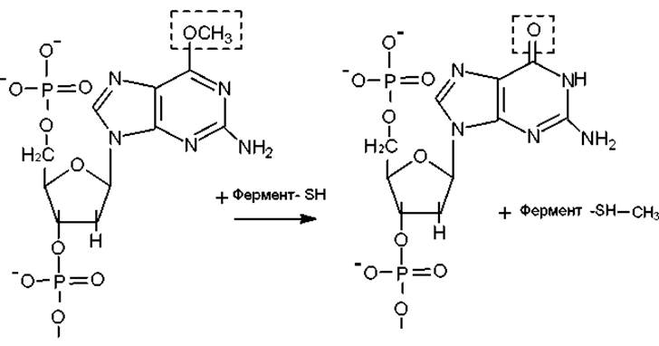
Fig. 2.3. The dealkylation of O6-methylguanine residues is catalyzed by a specific DNA alkyltransferase.
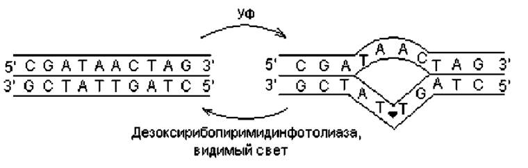
Fig. 2.4. Formation of thymine dimers induced by UV light and their light-dependent cleavage by a photoreactivating enzyme.
The phenomenon of photoreactivation is widespread in nature and has even been detected in such primitive microorganisms as Mycoplasmas. Photoreactivating enzymes have been found in certain Higher Plants and animals, as well as in all Bacteria studied, with the exception of Deinococcus radiodurans. Nevertheless, this bacterium is extremely resistant to UV light, surviving doses up to 1,000 times higher than those lethal to E. coli. Lacking photoreactivation entirely, D. radiodurans relies on a robust Excision Repair system.
Repair events associated with the replacement of distorted regions do not require visible light and involve, alongside Other Enzymes, two Major Types of Nucleases: exonucleases and endonucleases. Exonucleases degrade DNA starting from the strand ends, whereas endonucleases attack the internal Regions of the strands, generating single-stranded breaks in the DNA. Among The Diversity of repair pathways coupled with reparative DNA Synthesis, two main types can be distinguished: excision repair and post-replication repair.
Excision repair. A distinctive feature of excision repair is the removal of the damaged DNA segment. This type of repair is less specific to particular DNA lesions than photoreactivation and can correct not only pyrimidine dimers but also many other structural alterations in DNA. Excision repair (Fig. 2.5, A) is a multi-step process comprising the following events: 1) recognition of the DNA lesion, performed by specific endonucleases that also mediate the next stage; 2) incision—making a cut in One DNA strand near the lesion (carried out by endonucleases); 3) excision—removal of a nucleotide stretch containing the damage (carried out by exonucleases); 4) DNA resynthesis—filling the resulting gap (via DNA polymerase activity); 5) restoration of strand continuity through the formation of covalent bonds within the sugar-phosphate backbone.
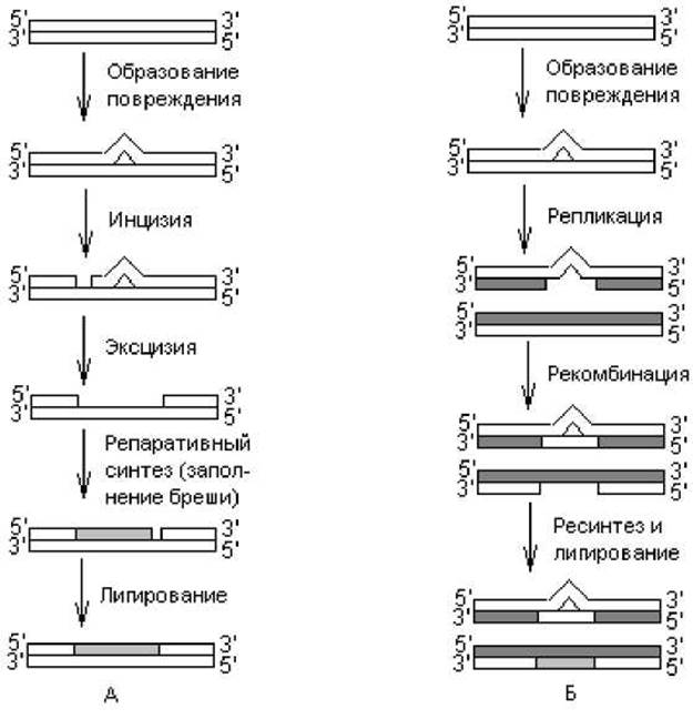
Fig. 2.5. Two Types of dark repair: A — excision repair, B — post-replication repair. Light lines represent original DNA strands; dark lines represent DNA strands synthesized during repair events.
The Mechanism of excision repair has been studied most thoroughly in the context of the dark removal of pyrimidine dimers from UV-irradiated E. coli DNA. In E. coli cells, this process is governed by the uvrA-D genes (which encode the enzymes that excise the DNA segment containing the dimer) and polA (which determines the structure of DNA polymerase I, responsible for reparative DNA synthesis). A hallmark of this excision repair pathway is the Introduction of single-stranded incisions on both sides of the thymine dimer.
Some organisms utilize yet another variant of excision repair to address damage, including that associated with thymine dimer formation, which involves a specialized enzyme known as N-glycosylase. In this case, the initial reparative event is the cleavage of the glycosidic bond between the damaged base (e.g., one of the thymines in a dimer, an N-alkylated purine, etc.) and deoxyribose. Thus, local apurinization or apyrimidinization occurs, generating a so-called AP site that is recognized by an AP-specific endonuclease, which then cleaves the phosphodiester bond adjacent to the AP site. The resulting gap is subsequently filled via standard repair synthesis.
A diverse array of N-glycosylases has been identified in both bacterial and Eukaryotic cells. For example, uracil-DNA glycosylase recognizes the mismatched dG/dU pair arising from the spontaneous deamination of a deoxycytosine residue in a dG/dC pair. Cytosine deamination can lead to the formation of a mutant dA/dT nucleotide pair during replication because, in terms of hydrogen bonding, uracil behaves similarly to thymine. Another widely distributed enzyme of this type is pyrimidine dimer-N-glycosylase, which generates an apyrimidinic site during the repair of Damage caused by pyrimidine dimer formation.
Sites that have undergone depurination or depyrimidination are excised by AP (apurinic and apyrimidinic) endonucleases. Both prokaryotic and eukaryotic cells possess a wide variety of AP endonucleases. Some of these enzymes nick the strand on the 3' side of the AP site, whereas others cleave the diester bond on the 5' side; in either case, 3'-hydroxyl and 5'-phosphoryl termini are generated. This enables an exonuclease to remove the adjacent residues on both sides of the nick along with the lesion.
Various Forms of excision repair are widespread among prokaryotic and eukaryotic organisms, including mammals. Impairments in excision repair pathways can lead to dramatic consequences. For instance, xeroderma pigmentosum is a well-known inherited human disorder whose primary symptom is extreme sensitivity to sunlight, ultimately leading to The Development of Skin Cancer. Various defects in excision repair have been identified in patients suffering from this condition.
Postreplication repair. This type of repair requires the participation of gene products that are also involved in recombination events (rec genes) and does not occur in rec mutant cells; hence, it is also referred to as recombinational repair. Recombinational postreplication repair relies on the replication and recombination of damaged DNA and is the least specific of all the repair mechanisms discussed, as it lacks a dedicated lesion-recognition step. It represents a relatively rapid pathway for restoring the native DNA Structure in daughter (newly synthesized) strands, with repair shown to take place within the first few minutes following irradiation. A distinctive feature of this process is that the lesion remains preserved in the original (parental) strands (Fig. 2.5, B).
Alongside this rapid pathway, a slow postreplication repair mechanism also exists, requiring several hours to complete. It is carried out by an enzyme system that is absent in unirradiated cells and is instead induced by irradiation. This mechanism is known as SOS repair. Its striking and paradoxical feature is a significant increase in the mutation frequency, even though the DNA is already damaged. This may be a consequence of utilizing a DNA strand containing lesions as a template.
Postreplication repair is present not only in bacteria but also in eukaryotic cells, including those of mammals.
Last update: 06/08/2026
Editorial and Educational Adaptation: This material has been compiled based on the primary/original source text. The project team performed an editorial review, corrected technical inaccuracies, structured sections, and adapted the content for an educational format.
What was processed:
- elimination of formatting defects (OCR errors, structural breaks, corrupted characters);
- editorial organization of content;
- standardization of terminology in accordance with academic sources;
- verification of factual statements against the original source text.
All mentions of the author, publication year, and origin of the primary text have been preserved in accordance with the source.