Amino Acids, Peptides and Proteins - Dévényi T., Gergely J. 1976
End-group analysis and stepwise degradation of proteins and peptides
Determination of N-terminal amino acids by the dansyl method
Amino groups of Proteins AND Peptides react with the fluorescent dye dimethylaminonaphthalenesulfonyl chloride (abbreviated as dansyl chloride) in an alkaline medium. As with dinitrophenylation, the bond between The amino acid and the reagent is not cleaved during Hydrolysis; therefore, DNS-Amino Acids can be identified in the hydrolysates of the dansylated protein (DNS-protein) or peptide. Such hydrolysates contain α-DNS-amino acids derived from N-terminal amino acids, as well as ε-DNS-Lys and O-DNS-Tyr derived from internal Lys and Tyr residues. In addition, dansyl chloride reacts with ammonia present in the solution to yield DNS-sulfonamide (DNS-NH2), while hydrolysis of the sulfonyl chloride group produces DNS-sulfonic acid (DNS-OH).
One advantage of Dansylation over dinitrophenylation is that following acid hydrolysis of the DNS-protein, the amino acids can be identified electrophoretically or chromatographically immediately after Protein Cleavage, without extracting the hydrolysate. Another advantage is its extremely high sensitivity. The fluorescence excitation maximum of DNS-amino acids is around 550 nm, and their fluorescence is so intense that paper Electrophoresis can easily detect 1–5 nmol, while Thin-Layer Chromatography can detect 0.2–1.0 nmol of the DNS derivative.
DANSYLATION TECHNIQUE [9]
Depending on the identification method, 0.3 to 5.0 nmol of the sample is introduced into a small test tube with an internal diameter of about 3–4 mm and a length of 25–30 mm. The solution volume is measured using a calibrated capillary. To avoid significant losses with small sample volumes (5–20 µl), any droplets of the test material should be spun down from the tube walls to the bottom by brief centrifugation at 2000–3000 rpm. The peptide solution is then dried in a vacuum desiccator; the dried sample is dissolved in 10 µl of 0.2 M NaHCO3 with gentle shaking and dried in vacuo again. The purpose of drying in this case is to remove ammonia, which interferes with the reaction. To the dry residue, 10 µl of deionized (ammonia-free) Water is added, dissolved with gentle shaking, and then 10 µl of a solution containing 2 mg/ml of DNS-Cl reagent in acetone is added. The mixture is gently shaken, the tube is sealed with Parafilm, and placed in an incubator at 37°C. Completion of the reaction is indicated by the disappearance of the yellow color, which usually occurs after half an hour. The reaction mixture is then dried, 30 µl of 6 N HCl is added, the tube is sealed, and the sample is hydrolyzed for 10–18 h at 105°C. After hydrolysis, the sample is centrifuged to bring any droplets down from the walls, then the tube is carefully opened and dried in vacuo over NaOH and P2O5.
IDENTIFICATION OF DNS-AMINO ACIDS
DNS-amino acids can be identified by paper or thin-layer electrophoresis, as well as by thin-layer chromatography.
IDENTIFICATION OF DNS-AMINO ACIDS BY PAPER ELECTROPHORESIS
To identify DNS-amino acids, electrophoresis must be performed several times. First, it is carried out in a pyridine-acetate buffer solution at pH 4.38 (Fig. 62). In this case, it is crucial that the pH is exactly 4.38, as a deviation of even 0.02 units significantly affects the Separation quality.
Class="center">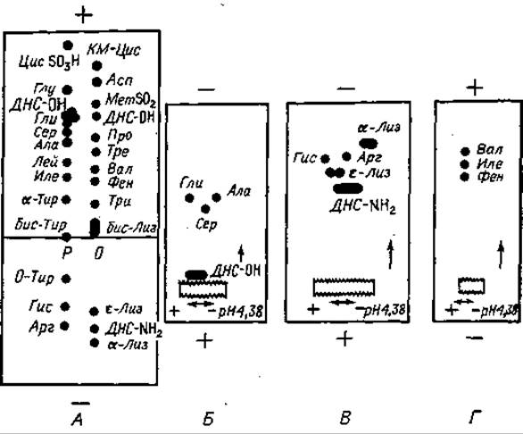
Fig. 62. Identification of DNS-amino acids by paper chromatography. A. Electrophoresis at pH 4.38. B, C, D. Electrophoresis at pH 1.9.
The buffer solution consists of 32 ml of acetic acid, 18 ml of pyridine, and 1950 ml of distilled water. The starting point is located at one-third of the sheet length from the cathode. The hydrolysate is applied to dry paper, keeping the starting spot as small as possible. Electrophoresis is run for 110 min at a voltage gradient of 80 V/cm. If high-voltage equipment is unavailable, intermediate voltage can be used by proportionally increasing the electrophoresis time. Direct liquid-cooled apparatus should not be used for the first electrophoresis, as DNS derivatives of Pro, Val, Ile, Leu, Phe, and Tyr can be extracted by organic Solvents.
Two control solutions of DNS-Amino acids are prepared separately. One solution (O) contains DNS-CM-Cys, DNS-Asp, DNS-MetSO2, DNS-Pro, DNS-Thr, DNS-Val, DNS-Phe, DNS-Trp, bis-DNS-Lys, α-DNS-Lys, ε-DNS-Lys, DNS-NH2. The other solution (P) contains DNS-Cys-SO3H, DNS-Glu, DNS-Gly, DNS-Ser, DNS-Ala, DNS-Leu, DNS-Ile, α-DNS-Tyr, DNS-His, DNS-Arg, O-DNS-Tyr, bis-DNS-Tyr. From these solutions, where the concentration of each derivative is 5 µmol/ml, 1 µl aliquots are taken for application.
After electrophoresis, the paper must be thoroughly dried in an oven at 100°C. Even a small amount of moisture or pyridine can cause severe fluorescence quenching, significantly reducing the sensitivity of the method. It is also recommended to place a Hair dryer behind the UV lamp to blow warm air onto the paper during visualization.
Sometimes, the separation of DNS-Gly, DNS-Ala, and DNS-Ser is unsatisfactory. The same applies to the group of DNS-Ile, DNS-Val, and DNS-Phe derivatives, and, in the presence of a strong DNS-NH2 spot, also to the group of DNS-His, DNS-Arg, α-DNS-Lys, and ε-DNS-Lys.
The identification of DNS-Gly, DNS-Ser, and DNS-Ala can be complicated by the accompanying DNS-OH spot; however, all four components are well resolved electrophoretically at pH 1.9. The section of the electrophoregram containing these four components (Fig. 62, B) is cut out, sewn onto another sheet of filter paper, and subjected to electrophoresis at pH 1.9. Fig. 62, B shows the distribution of derivatives obtained under these conditions. The zones containing basic DNS derivatives (Fig. 62, C) and DNS-Val, DNS-Ile, DNS-Phe (Fig. 62, D) are analyzed in the same manner.
If Val or Ile is at the N-terminus of the protein or peptide, the pattern may be complicated by the appearance of fluorescent peptides DNS-Val-X and DNS-Ile-X due to the resistance of Val-X and Ile-X peptide bonds to hydrolysis. The mobility of these derivatives differs from that of DNS-Val and DNS-Ile, which can lead to a false impression of heterogeneity in the studied peptide sample. In such cases, it is recommended to increase the hydrolysis time.
IDENTIFICATION OF DNS-AMINO ACIDS BY THIN-LAYER ELECTROPHORESIS [18]
DNS-amino acids can be quickly and reliably identified by thin-layer electrophoresis. Ready-to-use thin-layer electrophoresis plates are currently commercially available (e.g., MN-Polygram, Sil N-HR, Macherey-Nagel Co., FRG).
On a 20 x 10 cm plate, the DNS-peptide hydrolysate is applied to the starting point marked 8 cm from the cathode, with continuous drying. The control amino acid mixtures have the following composition. Solution K: DNS-Cys-SO3H, DNS-Asp, DNS-Ser, DNS-Thr, DNS-Pro, α-DNS-Tyr, DNS-Phe, bis-DNS-Lys, DNS-Arg, DNS-Leu, ε-DNS-Lys, α-DNS-Lys. Solution L: DNS-CM-Cys, DNS-Glu, DNS-Gly, DNS-Ala, DNS-Met, DNS-Val, bis-DNS-Tyr, O-DNS-Tyr, DNS-Ile, DNS-His.
The buffer solution contains 12 ml of acetic acid and 8 ml of pyridine in a final volume of 1 L (pH 4.5). Wetting of the plate is performed as follows: the buffer solution is poured into a Glass chamber or a photographic tray, and the plate is carefully immersed in it. When the solution level reaches the starting line, the plate is removed, blotted to remove excess solution, and then immersed back into the solution from the opposite side. The starting line itself is wetted only when both liquid fronts meet; this avoids direct wetting of the starting line.
Electrophoresis is carried out in a horizontal apparatus at a voltage gradient of 80 V/cm. The current for a 10 cm plate width should be 50–60 mA. After 20–25 min of electrophoresis, the plate is removed, dried with a stream of warm air, and the DNS-amino acids are identified under UV light.
The electrophoregram is shown in Fig. 63. The separation of Phe, Ile, and Leu may present certain difficulties, as does the identification of basic DNS-Amino Acids and the separation of bis-DNS-Lys and bis-DNS-Tyr. To separate Phe, Ile, and Leu, one should first perform electrophoresis on another plate, followed by chromatography in the perpendicular direction in a benzene–pyridine–acetic acid (80 : 20 : 2) system. By this procedure, DNS-Phe is identified with absolute certainty, and DNS-Ile and DNS-Leu are resolved very clearly. DNS-His is determined using the Pauly reaction immediately after electrophoresis; in this way, it is differentiated from other basic Amino Acid Derivatives: DNS-Lys and DNS-Arg.
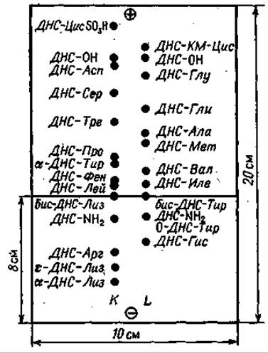
Fig. 63. Identification of DNS-amino acids by thin-layer electrophoresis at pH 4.5 in a pyridine–acetic acid mixture.
IDENTIFICATION OF DNS-AMINO ACIDS BY THIN-LAYER CHROMATOGRAPHY ON POLYAMIDE PLATES [18]
All DNS-amino acids can be determined with a high degree of reliability by thin-layer chromatography on polyamide plates. Identification is facilitated by the "sandwich" design of the polyamide plates, i.e., both sides of the support plate are coated with a thin layer of polyamide. The DNS-amino acid under study is chromatographed on one side of the plate, while a reference mixture of amino acids is applied to the other. Taking advantage of the plate's transparency, the unknown DNS-amino acid can be identified after chromatography.
The sequence of operations in this method is as follows. In one of the corners of the plate, at a distance of 1 cm from the edges, a starting point is marked. Care must be taken to ensure that this point is in the exact same position on both sides. The hydrolysate, prepared as described above, is then dissolved in 20 µl of 50% pyridine; half of it is applied to one side and half to the other, drying the spot continuously. It is important to apply the material strictly as a single spot. Next, a reference mixture of amino acids is applied to one side of the plate, consisting of the following components: DNS-Ser, DNS-Glu, DNS-Pro, DNS-Gly, DNS-Ile, bis-DNS-Tyr, and DNS-Arg. In the reference mixture, 1 µl of which is applied to the starting point, the concentration of each derivative is 1 nmol/µl.
Generally, to identify the amino acids, chromatography must be performed in three solvent systems: I — 1.5% formic acid; II — benzene–acetic acid (9 : 1); III — ethyl acetate–methanol–acetic acid (10 : 1 : 1). In some cases, for the identification of ε-DNS-Lys, α-DNS-His, and DNS-Arg, a fourth system may be required: IV — 0.05 M Na3PO4 — ethanol (3 : 1). For the identification of DNS-cysteic acid, a fifth solvent system is required: V — 1 M NH4OH — ethanol (1 : 1).
The test samples are first chromatographed in solvent I for 30 min, and then, after drying the plate with a hair dryer, for 60 min in the perpendicular direction in system II. The plate is dried again, and the separation of the components is checked under ultraviolet light. Chromatography in system III (in the same direction as in system II) is carried out only when it is necessary to identify DNS-Thr and DNS-Ser, or DNS-Asp and DNS-Glu. In this case, chromatography is continued for 45 min. If DNS-Cys SO3H is present in the mixture, then after chromatography in system II, the samples are chromatographed for 40 min in the same direction in system V. To separate ε-NH2-DNS-Lys, α-NH2-DNS-His, and DNS-Arg, after chromatography of the mixture in system II, it is chromatographed for 45 min in system IV.
The distribution of spots on the chromatogram is shown in Fig. 64. Polyamide plates can be reused several times if they are thoroughly washed after each run as follows: the plate is immersed in 50% acetone containing 1 M NH4OH, taking care not to Touch the walls of the vessel, and washed for 2 h with constant stirring of the washing solution. After drying, the plates can be used again, and it is preferable to maintain the same direction of solvent flow.
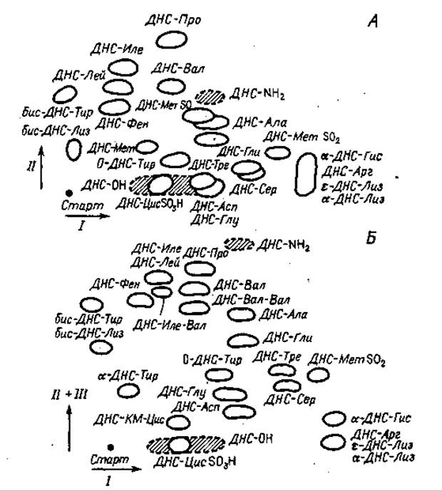
Fig. 64. Identification of DNS-amino acids by thin-layer chromatography on polyamide.
Solvents: I — 1.5% formic acid, II — benzene–acetic acid (9:1), III — ethyl acetate–methanol–acetic acid (10:1:1, by volume). Abbreviations: CM-Cys — S-carboxymethylcysteine; Cys SO3H — cysteic acid, Met SO — Methionine sulfoxide, Met SO2 — methionine sulfone.
DNS-Arg is determined by the Sakaguchi reaction. Bis-DNS-Lys and bis-DNS-Tyr can be separated by chromatography In the second direction using the system 2-butanone–propionic acid–water (15 : 5 : 6).
The DNS derivative of the N-terminal Trp is completely destroyed during hydrolysis. Therefore, a completely negative result in amino acid dansylation experiments may indicate that Trp is present at the N-terminus. DNS-Trp can be easily identified after chymotryptic hydrolysis. All three Tyr derivatives (bis-DNS-Tyr, O-DNS-Tyr, and α-DNS-Tyr) are usually easily identified; however, due to its poor solubility in water, the bis-derivative is often not detected, even though it is known to be present in the mixture. In such cases, the hydrolysate is dissolved in pyridine and applied to paper or a thin-layer plate. DNS-CM-Cys is characterized by particularly high losses during hydrolysis, so more material should be used for analysis if this amino acid is expected to be at the peptide terminus.
The DNS-amino acids required for reference solutions do not always need to be isolated in pure crystalline form. It is quite sufficient to dansylate the amino acid solution, as described below, without isolating them from the solution. By-products such as DNS-OH and DNS-NH2 do not interfere with identification and therefore do not need to be removed from the mixture.
The isolation method described below for DNS-Ala can also be used to obtain the following homogeneous crystalline derivatives: DNS-Gly, DNS-Val, DNS-Leu, DNS-Ile, DNS-Phe, DNS-Ser, DNS-Thr, DNS-Pro, DNS-Met, DNS-Met SO2, DNS-Cys, DNS-Asp, and DNS-Glu.
150 mg of Alanine is dissolved in 5 ml of 1 M sodium bicarbonate. To this solution, 100 mg of DNS-Cl dissolved in 5 ml of acetone is added with vigorous stirring. If some of the DNS-Cl precipitates, it is dissolved by adding 0.2 ml of trimethylamine. After stirring for 10 min, the reaction mixture is extracted with butyl acetate (3 times with 7 ml) to remove acetone, DNS-NH2, and unreacted DNS-Cl. The pH of the aqueous phase is adjusted to 3.4 with 2 N HCl, and DNS-Ala is extracted with benzene (3 times with 5 ml). Piperidine is carefully added dropwise to the benzene extract until turbidity appears, and the mixture is left in the cold. Recrystallization is carried out from butyl acetate.
Last update: 19/08/2026
Editorial and Educational Adaptation: This material has been compiled based on the primary/original source text. The project team performed an editorial review, corrected technical inaccuracies, structured sections, and adapted the content for an educational format.
What was processed:
- elimination of formatting defects (OCR errors, structural breaks, corrupted characters);
- editorial organization of content;
- standardization of terminology in accordance with academic sources;
- verification of factual statements against the original source text.
All mentions of the author, publication year, and origin of the primary text have been preserved in accordance with the source.