Biological Membranes - A. N. Ogurtsov 2012
Electrogenesis of Biomembranes
Membrane Receptors
Tyrosine Kinase Receptors and MAP Kinase Cascades
ADP-stimulated activation of Blood Coagulation (Figure 139) also triggers regulated exocytosis of specific Proteins by platelets, namely platelet-derived growth factors (PDGF). These growth factors initiate the regeneration of damaged Blood Vessels.
Blood vessel Cells contain PDGF receptors within their membranes. The PDGF molecules bind to the exoplasmic domains of these receptors, while their cytosolic domains possess Tyrosine kinase activity. This means that upon activation by PDGF binding, the cytosolic domain of the receptor can phosphorylate tyrosines on other proteins.
A single platelet-derived growth factor molecule can bind to two closely spaced PDGF receptors, causing them to mutually phosphorylate each other—a process known as autophosphorylation (Figure 147).
The Structure of many cytosolic proteins features a so-called SH2 domain, at the bottom of whose deep pocket a positively charged Arginine residue is located. The SH2 domain is designed to bind phosphorylated tyrosine. Other Phosphorylated Amino Acids simply cannot reach the bottom of the pocket; consequently, their binding affinity is insufficient to form a stable protein-protein complex.
Class="center">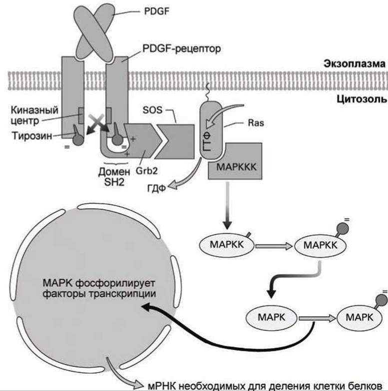
Figure 147 - Diagram of the MAP kinase cascade initiation
Therefore, proteins containing an SH2 domain bind to the dimerized PDGF receptor, but they do not bind to a monomeric PDGF receptor that lacks the phosphorylated tyrosine containing the negatively charged phosphate group necessary for binding.
One of the proteins possessing an SH2 domain is growth factor receptor-bound protein 2 (Grb2). Via Grb2, another protein, SOS, attaches to the activated PDGF receptor, and in this complex, the SOS protein acts as a guanine nucleotide exchange factor (GEF) for a GTPase called Ras. As a result, the GEF protein SOS stimulates the exchange of GDP for GTP in the Ras protein, switching it to the active state.
The activated Ras protein triggers an enzymatic cascade consisting of protein Kinases that phosphorylate one another and, at the end of the cascade, activate (via phosphorylation) the final protein kinase known as mitogen-activated protein kinase or MAP kinase (MAPK).
The naming of the kinases that make up this cascade lacks originality. The kinase that phosphorylates MAP kinase is called MAP kinase kinase or MEK (MAPKK). In turn, it is phosphorylated by MAP kinase kinase kinase or Raf (MAPKKK). And it is this "third" level kinase that is activated by the Ras protein (see [1], sec. 6.8.4).
The word "mitogen" in the name of these kinases indicates that they are involved in stimulating The process of mitosis (MITosis GENeration, mitogen); thus, the MAP kinase activated in this complex manner by PDGF triggers Cell Division.
Phosphorylated MAP kinase is translocated into The Nucleus, where it phosphorylates METABOLISM/31.html">Transcription factors of those genes whose expression culminates in DNA Replication and the synthesis of proteins required for mitosis.
Platelet-derived growth factor is just one of many growth factors, all of which initiate similar cascades. First, the growth factor binds to a dimerized tyrosine kinase receptor (specific to each factor). Next, an SH2 domain-containing protein (such as Grb2) attaches to the phosphorylated tyrosine residues of the receptors. This activates the Ras protein and the entire MAP kinase cascade, leading to DNA replication and cell division.
It is worth noting an important therapeutic application involving MAP kinase cascades. These cascades are turned off when the GTPase Ras is deactivated through GTP Hydrolysis.
A mutant form of the Ras protein with impaired GTPase activity fails to hydrolyze GTP, does not undergo deactivation, and consequently chronically activates the MAP kinase cascade, leading to cell proliferation even in the absence of growth factors.
Such mutant forms of the Ras protein are found in 20% of human Cancer cells.
The drug R115777 blocks the phosphorylation of the MAPKKK kinase by the Ras protein, interrupting the cascade and halting the uncontrolled cell division characteristic of oncological diseases.
In addition to MAP kinase cascades, growth factors can stimulate the release of Calcium Ions into the Cytosol from The Endoplasmic reticulum. Phosphoinositide phospholipase C gamma isoform (PLCγ) also contains an SH2 domain, and its binding to a tyrosine kinase receptor leads to its phosphorylation (activation) (Figure 148).

Figure 148 - Diagram of phospholipase C γ activation
Active PLCγ hydrolyzes PIP2, and the resulting Inositol trisphosphate triggers the release of calcium from the endoplasmic reticulum.
Last update: 13/08/2026
Editorial and Educational Adaptation: This material has been compiled based on the primary/original source text. The project team performed an editorial review, corrected technical inaccuracies, structured sections, and adapted the content for an educational format.
What was processed:
- elimination of formatting defects (OCR errors, structural breaks, corrupted characters);
- editorial organization of content;
- standardization of terminology in accordance with academic sources;
- verification of factual statements against the original source text.
All mentions of the author, publication year, and origin of the primary text have been preserved in accordance with the source.