Genetics - A. V. Sivolob 2008
The Nature of Genetic Material
Cell Cycle and Cell Division in Eukaryotes
METABOLISM/36.html">DNA Replication occurs during the so-called S phase of the Cell Cycle, which, in turn, is a stage of interphase—the period when Chromosomes exist as Chromatin fibrils and The Cell expresses Genetic information (Fig. 1.30). Following replication and its accompanying repair processes, sister chromatids (future daughter chromosomes) generated by DNA duplication remain connected through interaction with specific Proteins. The cell then enters the G2 phase, which can be viewed as preparation for mitosis, the Cell Division of somatic Cells.
Class="center">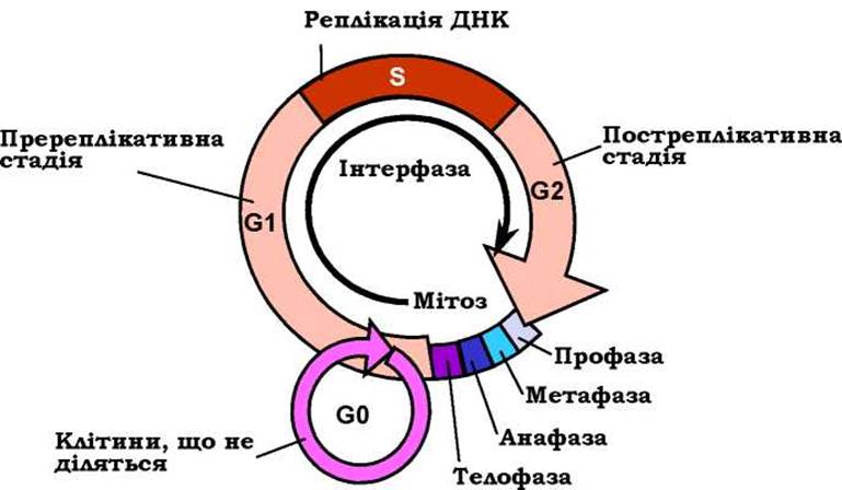
Fig. 1.30. Scheme of the cell cycle
During the first stage of mitosis—prophase—chromatid Condensation takes place, alongside the maturation of two centrosomes (structures consisting of two centrioles each), The formation of the mitotic spindle, and the attachment of spindle fibers to the chromosomes (Fig. 1.31). Toward the end of prophase, the centrosomes migrate toward opposite poles of the cell.

Fig. 1.31. Scheme of cell division via mitosis.
Two pairs of homologous chromosomes are shown
Next, in metaphase, chromosomes (pairs of sister chromatids joined at their centromeres) align along the cell equator. Anaphase begins with the Separation of centromeres, after which the sister chromatids, now referred to as daughter chromosomes, move toward the poles. During telophase, nuclear membrane formation and chromosome decondensation begin. Telophase (and mitosis itself) concludes with cytokinesis—the division into two daughter cells.
Upon completion of mitosis, a cell destined for further division enters the G1 phase of the cell cycle (which can be considered pre-replicative), whereas a terminally differentiated cell for which this was the final mitosis enters the G0 phase (Fig. 1.30). The outcome of mitosis is two identical cells with two identical diploid chromosome sets.
During the formation of Germ Cells (Gametes) via Meiosis in a diploid precursor cell, DNA Replication and chromatid duplication also occur, with the chromatids remaining linked by their centromeres.
Meiosis proceeds through two successive cell divisions (Fig. 1.32). In prophase I (which encompasses the leptotene, zygotene, pachytene, diplotene, and diakinesis stages—not detailed in the figure), so-called bivalents, or tetrads, are formed as complexes of homologous chromatid pairs. Two of the four chromatids of a bivalent, belonging to homologous chromosomes, form synaptonemal complexes at several points with the participation of specific proteins, where Homologous Recombination begins (Fig. 1.32). As a result, segments are exchanged between homologous chromosomes. This is followed by the gradual condensation of chromatids, revealing characteristic cross-like structures between them—chiasmas—formed as a consequence of recombination. Prophase I ends with the separation of the condensed bivalents. Subsequently, in metaphase I, the chromosome pairs align along the equator; in anaphase I, they segregate; and in telophase I, the two cells containing a double set of DNA molecules finally divide. Meanwhile, the sister chromatids remain connected at their centromere regions.

Fig. 1.32. Scheme of cell division via meiosis.
One pair of homologous chromosomes is shown
Next, during the Second Division (prophase II, metaphase II, anaphase II, and telophase II are not shown in Fig. 1.32), centromeres split, chromosomes segregate, and both cells divide, resulting in four haploid gametes. Upon Fertilization, the parental chromosome sets combine to form a diploid zygote, which subsequently gives rise to all other Cells of the multicellular offspring Organism through mitotic division.
Last update: 11/08/2026
Editorial and Educational Adaptation: This material has been compiled based on the primary/original source text. The project team performed an editorial review, corrected technical inaccuracies, structured sections, and adapted the content for an educational format.
What was processed:
- elimination of formatting defects (OCR errors, structural breaks, corrupted characters);
- editorial organization of content;
- standardization of terminology in accordance with academic sources;
- verification of factual statements against the original source text.
All mentions of the author, publication year, and origin of the primary text have been preserved in accordance with the source.