Study Guide on Tuberculosis - M.M. Savula 2002
Diagnostic Methods for Tuberculosis
Radiological Diagnosis
Radiological examination Methods are among the primary tools for diagnosing tuberculosis in various locations, especially the Lungs.
Fluoroscopy. This involves directly examining the patient under an X-ray screen. This method allows for rapid results, examination of the patient in various projections, and Assessment of the function of different Organs (respiratory depth, cardiac pulsation). During fluoroscopy, a site for pleural puncture can be marked, and Procedures like angiocardiography and bronchography can be monitored. Equipment with electronic-optical image intensification is used.
Radiography. This involves taking an X-ray image in direct (AP/PA) and lateral projections. The examination can be repeated to monitor the process's dynamics, and the image can be reviewed by a qualified specialist.
Fluorography is used for mass population screenings. The throughput of a fluorograph is up to 50-60 people per hour. It is less expensive than radiography. The diagnostic value of a high-quality fluorogram approaches that of a radiograph, and it can be performed in various projections. Therefore, in recent times, fluorography has also been utilized as a diagnostic method.
In recent years, low-dose digital fluorographs have been developed and implemented, significantly reducing the radiation exposure to the patient. They allow for the detection of small contrast-enhancing lesions and simultaneous examination of The Structure of the lung parenchyma, Trachea, large Bronchi, vertebral bones, and chest wall.
Analysis of Chest Radiographs and Fluorograms Performed in the Posteroanterior (PA) Projection
To properly evaluate a radiograph or fluorogram, it must be performed correctly. The patient stands straight, is asked to stand close to the screen or cassette, place their hands on their hips, turn their elbows forward (to prevent scapular shadows from overlapping the lung fields), take a deep breath, and hold it. The image is captured at the peak of inspiration.
Radiographs are viewed on a light box (negatoscope), while fluorograms are examined under the magnifying lenses of a fluoroscope.
Air-filled lung tissue allows X-rays to pass through, whereas dense Tissues and fluid (Heart, Blood Vessels, bones, inflamed or necrotic lung tissue, effusion) attenuate X-rays and produce a corresponding image on the X-ray film. In practice, a negative image on film is used, where all dense structures appear bright, and air-filled lung tissue appears dark but translucent. Therefore, bright areas against the lung parenchyma are conventionally referred to as opacities or shadows, while dark areas are called lucencies. A normal chest radiograph in the posteroanterior (PA) projection is shown in Fig. 4. On the chest radiograph, the central position (slightly more to the left) is occupied by the mediastinal shadow (of The Heart and great vessels) (1). On both sides of the mediastinal shadow, translucent lung fields are visible, bounded inferiorly by the diaphragmatic domes (2). The right hemidiaphragm is positioned slightly higher than the left. The lung fields are traversed by arc-shaped rib shadows. Their posterior segments (3) are somewhat more intense, while the anterior segments (4) are brighter.
The cartilaginous PARTS OF THE Ribs are not visible because X-rays penetrate them more easily, and anteriorly, the ends of the ribs appear to disappear before reaching the Sternum.
The area of lung tissue above the clavicle (5) is referred to as the apex. Lung tissue on an X-ray image has a specific structure.
On both sides of the cardiovascular shadow, the shadows of the lung hila (6) are visible. Their shadow begins superiorly at the level of the II- III ribs and extends inferiorly for two intercostal spaces (to the IV- V ribs). In terms of their
shape, the right hilum resembles a comma, while the left, positioned slightly higher, is semi-oval. The anatomical substrate of the lung hilum includes large arterial and venous vessels through which blood flows to and from the heart, bronchi, lymphatic nodes and vessels, nerve trunks, and Connective Tissue. However, the hilar shadow visible on the radiograph is primarily caused by blood-filled vessels, which are poorly permeable to X-rays. Shadows of hilar Lymph Nodes become visible only in cases of their pathological enlargement due to various diseases. Due to its heterogeneous Anatomical Structure, the hilar shadow is normally non-uniform.
Class="center">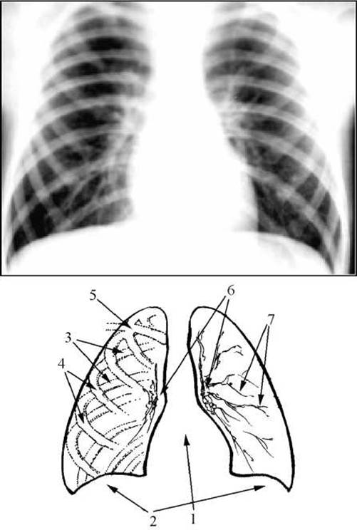
Fig. 4. Normal chest radiograph.
Striated shadows radiate from the hilar shadow, dividing dichotomously and gradually tapering towards the periphery. The totality of these radial striated shadows is referred to as the pulmonary vascular pattern (7). Their primary anatomical substrate consists of the branching pulmonary Arteries and Pulmonary Veins.
To evaluate changes on a radiograph, a General Overview is first performed to ascertain whether all lung regions are included (apices, Diaphragm), whether the lung fields are symmetrical, and if the cardiac shadow occupies a central position. The structure and position of the hila and the pulmonary vascular pattern are assessed. Subsequently, symmetrical lung segments are systematically examined from top to bottom, searching for pathological shadows. In Pulmonary Tuberculosis, four types of shadows are most commonly detected:
- foci - shadows up to 1 cm in size (Fig. 5);
- infiltrates - all shadows larger than 1 cm, which may occupy even a segment, lobe of the lung, or more (Fig. 6); if a lucency appears in the center of an infiltrate, this indicates infiltrate breakdown and cavity formation;
- ring-shaped shadows - a ring completely closed along its entire perimeter, representing the radiological manifestation of a pulmonary cavity - a cavern, abscess, or air cyst (Fig. 7);
- linear shadows represent scar strands, or adhesions.

Fig. 5. Chest radiograph. A group of intense foci is visible below the right clavicle.
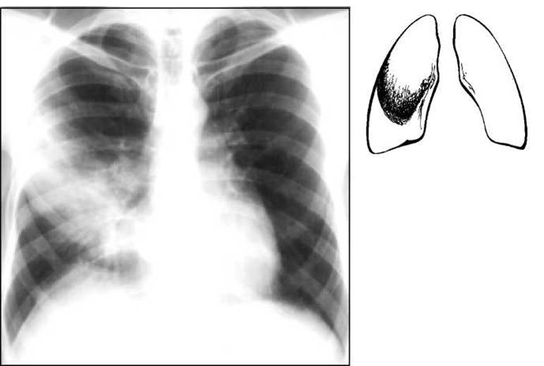
Fig. 6. Chest radiograph. Infiltrative shadow in the right lung.
The most characteristic feature of pulmonary tuberculosis is the presence of focal lesions, which accompany all forms of the disease. These may be the sole manifestation of pulmonary tuberculosis or may be found alongside infiltrates and ring-shaped shadows. The presence of focal shadows in the lungs sometimes allows for the differentiation of tuberculosis from other pulmonary diseases.
In Primary tuberculosis in children, the hilar lymph nodes are typically affected and enlarged. On a radiograph, this manifests as hilar enlargement and the appearance of additional rounded, sometimes polycyclic shadows of the enlarged lymph nodes (Fig. 8).
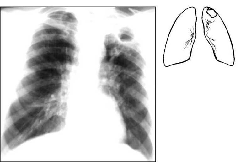
Fig. 7. Chest radiograph. A ring-shaped shadow (cavity) is visible at the left apex, with focal lesions below.
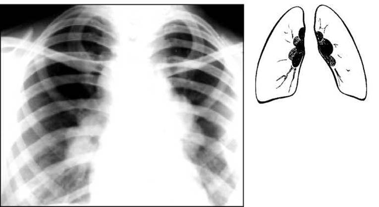
Fig. 8. Chest radiograph. Polycyclic shadows of enlarged Lymph Nodes in the hilar regions.
Tomography is a sectional study performed at various depths (Fig. 9), allowing for a more precise Determination of the structure (particularly the presence of small cavities) and localization of a pathological formation. Tomograms eliminate the bony framework of the chest, which might otherwise obscure pathological shadows. Tomography is performed with the patient in a supine position. To examine Changes in the hilar LYMPH NODES AND bronchial patency, a so-called median slice is taken—calculated as half the distance from the table to The surface of the sternum at the height of inspiration.
Computed tomography (CT) provides images of cross-sections of The Human Body at various levels (Fig. 10). It can differentiate between tissues and formations with slight differences in density, allow for a detailed study of their structure, and detect small cavities, slightly enlarged hilar lymph nodes, as well as mesenteric and retroperitoneal lymph nodes. It is also used for the Differential Diagnosis between Tuberculous meningitis and Brain Tumors.

Fig. 9. Tomogram of the right lung at a depth of 7 cm. The upper lobe is reduced in volume and unevenly opacified. The trachea is shifted to the right.

Fig. 10. Computed tomography scan.
Bronchography (Fig. 11) involves the targeted Filling of the bronchi with a contrast medium (such as sulfoyodolipol or yodolipol) following local anesthesia, followed by X-rays in two projections. This method can detect bronchial narrowing, deformations, stenoses, and Bronchiectasis.
Angiopulmonography and bronchial arteriography are contrast-enhanced methods used to examine the pulmonary vessels. These procedures are performed in specialized thoracic departments to detect pulmonary vessel thrombosis and developmental anomalies.
Last update: 10/08/2026
Editorial and Educational Adaptation: This material has been compiled based on the primary/original source text. The project team performed an editorial review, corrected technical inaccuracies, structured sections, and adapted the content for an educational format.
What was processed:
- elimination of formatting defects (OCR errors, structural breaks, corrupted characters);
- editorial organization of content;
- standardization of terminology in accordance with academic sources;
- verification of factual statements against the original source text.
All mentions of the author, publication year, and origin of the primary text have been preserved in accordance with the source.