Tuberculosis - I. T. Pyatnochka 2005
Complications of tuberculosis
Spontaneous pneumothorax
Spontaneous pneumothorax refers to the partial or complete collapse of a lung caused by The entry of air into the pleural cavity due to a breach in the integrity of the visceral Pleura (Fig. 32).
Among all Complications of Pulmonary Tuberculosis, spontaneous pneumothorax accounts for no more than 1–2%.
Pathogenesis. Spontaneous pneumothorax most commonly occurs in individuals with chronic pulmonary conditions, including tuberculosis, As a result of the rupture of subpleural bullae in vicarious emphysema, and very rarely due to the rupture of a cavity wall. The triggering factor for spontaneous pneumothorax is an increase in intrapulmonary pressure, particularly within the area of thin-walled bullae, during physical exertion, coughing, etc.
The Clinical presentation of spontaneous pneumothorax depends on the rate at which air enters and its volume in the pleural cavity. It is classified into total (complete lung collapse) and partial (lung collapse by 1/3–1/2); depending on the pressure within the pleural cavity, it is categorized as closed, open, or tension (valvular) spontaneous pneumothorax. The specific type of spontaneous pneumothorax is determined by manometry: in a closed pneumothorax, intrapleural pressure is lower than atmospheric pressure (-), in an open pneumothorax, it equals atmospheric pressure (±), and in a tension pneumothorax, it is higher (+) than atmospheric pressure. The clinical picture depends on The rate of development, degree of collapse, mediastinal shift, and the functional state of the cardiopulmonary system. A particularly severe and life-threatening form of spontaneous pneumothorax is tension pneumothorax, in which The amount of air in the pleural cavity increases with each inhalation, leading to rising intrapleural pressure, progressive lung collapse, and a shift of the mediastinal Organs to the opposite side. Clinically, this manifests as signs of Acute Respiratory Failure with hemodynamic disturbances, and occasionally as pleural Shock with loss of consciousness, which can be fatal if therapeutic measures are not taken. Closed spontaneous pneumothorax has a relatively mild clinical course.
Diagnosis is based on medical history—predominantly a chronic pulmonary disease. Spontaneous pneumothorax often develops acutely, triggered by precipitating factors (such as a sharp cough or physical exertion), and is accompanied by side pain, dyspnea, coughing, and occasionally cyanosis. The chest on the affected side is enlarged and significantly lags behind during respiratory movements. Palpation reveals absent vocal fremitus, Percussion reveals tympanitic Resonance, and Auscultation shows decreased or absent breath sounds. Subcutaneous emphysema may occasionally be present. However, the most informative diagnostic method is radiography (clearly showing the edge of the collapsed lung, beyond which the pulmonary pattern is absent, frequently with a horizontal fluid level). Computed tomography is highly valuable in the Cytology/practical/136.html">Differential diagnosis OF spontaneous pneumothorax, cysts, or a large, inflated, thin-walled bulla. Thoracoscopy helps clarify A number of specific details regarding spontaneous pneumothorax.
Class="center">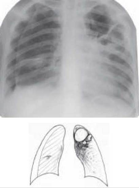
Fig. 32. Right-sided spontaneous pneumothorax. Fibrocavernous tuberculosis of the left lung
Differential diagnosis of spontaneous pneumothorax is performed to distinguish it from traumatic and artificial pneumothorax, giant cavities, air cysts, and large bullae.
Treatment is carried out in a hospital Setting, prescribing symptomatic medications (sulfocamphocain 10% – 2 ml subcutaneously, cordiamine 2 ml intramuscularly or subcutaneously, Oxygen therapy); antitussives (dionine – 0.015 g twice daily orally, codterpin 1 tab 2–3 times daily orally, ambroxol (lasolvan) 0.03 g two to three times daily orally); analgesics (omnopopon or promedol 1–2% – 1 ml subcutaneously, analgin 50% – 2 ml intramuscularly, baralgin 5 ml intramuscularly). In cases of minor lung collapse and satisfactory General condition of the patient, bed rest is prescribed for 7–10 days. If the volume of air exceeds 1/3 of the hemithorax, periodic pleural punctures and air aspiration are performed. If punctures are ineffective, a tube is inserted into the pleural cavity for continuous air aspiration. Occasionally, endoscopic occlusion of the affected segmental or lobar bronchus using a hemostatic (or less commonly, поролонова / polyurethane foam) sponge can be used to achieve aerostasis. If conservative treatment yields no positive outcome, the question of thoracotomy and suturing of the injured lung or its resection is considered.
In tension (valvular) pneumothorax, emergency relief can be achieved by inserting a large-bore needle or trocar into the pleural cavity while the patient is in a semi-sitting position. If this Procedure fails to reduce intrapleural pressure and does not eliminate the immediate threat to the patient's life, pleural drainage is performed using the Bülau method: a plastic catheter is inserted into the pleural cavity via a trocar, fixed to the Skin with a ligature, and the distal end of the catheter—fitted with a cut finger from a rubber glove—is immersed in a Glass jar containing furatsilin. The patient is then transferred to a specialized medical facility.
Last update: 10/08/2026
Editorial and Educational Adaptation: This material has been compiled based on the primary/original source text. The project team performed an editorial review, corrected technical inaccuracies, structured sections, and adapted the content for an educational format.
What was processed:
- elimination of formatting defects (OCR errors, structural breaks, corrupted characters);
- editorial organization of content;
- standardization of terminology in accordance with academic sources;
- verification of factual statements against the original source text.
All mentions of the author, publication year, and origin of the primary text have been preserved in accordance with the source.