Review of Medical Physiology - William F. Ganong 2002
Physiology of Nerve and Muscle Cells
Excitable Tissue: Muscle
Cardiac Muscle (Myocardium) - Mechanical Properties
Contractility
The contractile response of the myocardium begins immediately after the onset of depolarization and lasts nearly one and a half times longer than the Action Potential (see Fig. 3-14). The Role of Ca2+ in coupling excitation with contraction is the same as in Skeletal Muscle (see above). However, as noted earlier, it is the actual influx of Ca2+ into The Cell from the extracellular fluid—driven by the activation of dihydropyridine channels in the T-system—rather than depolarization itself that triggers the release of Ca2+ ions from their storage sites in the sarcoplasmic reticulum.
Class="center">
Fig. 3-13. A: Electron micrograph of cardiac muscle. The indistinct thick lines represent intercalated discs (magnification 12,000×) (reproduced with permission from Bloom W, Fawcett DW: A Textbook of Histology, 10th ed. Saunders, 1975). B: Schematic diagram of cardiac muscle under light and Electron Microscopy; N - Nucleus (reproduced with permission from Braunwald E, Ross J, Sonnenblick EH: Mechanisms of contraction of the normal and failing Heart. N Engl J Med 1967;277:794. Courtesy of Little, Brown, Inc.)
During phases 0-2 and for about half of phase 3 (until the Membrane Potential reaches approximately -50 mV during repolarization), cardiac muscle cannot be re-
excited; in other words, it is in a state of absolute refractory period (see Fig. 3-4). It remains relatively refractory until the onset of phase 4. Consequently, tetanus of the type observed in skeletal muscle is impossible in cardiac muscle. Obviously, even a brief transition of the myocardium into a tetanic state could have fatal consequences, and from this perspective, the inability of cardiac muscle to undergo tetanus serves as a safety mechanism.
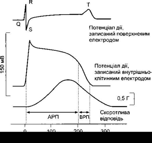
Fig. 3-14. Action potentials and contractile responses of mammalian cardiac muscle represented as potential-time curves; ARP - absolute refractory period; RRP - relative refractory period.
Isoforms
Cardiac muscle is generally slow and exhibits a relatively low ATPase activity. Its function relies on oxidative METABOLISM and, consequently, on a continuous supply of O2. Human cardiac muscle contains both α- and β-isoforms of Myosin heavy chains (α-MHC and β-MHC). The β-MHC chain possesses lower myosin ATPase activity than the α-MHC. Both isoforms are present in the atrium, where the α-isoform predominates, whereas only the β-isoform is found in the ventricle. The Influence of THYROID Hormones on MHC isoforms is discussed in Chapter 18. Thyroid hormones also induce changes in skeletal muscle MHC isoforms, although in this case, their effect is more complex and multi-component.
Relationship Between Muscle Fiber Length and Developed Tension
The relationship between the initial length of a muscle fiber and the total tension developed in cardiac muscle is identical to that observed in skeletal muscle. There is an optimal resting length at which the tension generated in response to external stimulation is maximal. Initial fiber length is determined by the diastolic Filling of the heart chambers, and the pressure developed within the ventricle is proportional to its total tension (Starling's law of The Heart, see Chapter 29). Consequently, the force of contraction (Fig. 3-16) increases with an increase in diastolic volume until it reaches a maximum (the ascending limb of Starling's curve), after which it begins to decline (the descending limb of Starling's curve). However, unlike skeletal muscle, the reduction in contractile force during significant stretching is not caused by a decrease in the number of Actin-myosin cross-bridges, because even with marked chamber dilation, stretching of the myocardium to such an extent does not occur in vivo. The descending limb of Starling's curve corresponds to the initial structural damage of cardiac fibers. The homeostatic significance of Starling's law is discussed in Chapter 29.

Fig. 3-15. Top: Phases of The cardiac muscle fiber action potential: 0 - depolarization; 1 - initial rapid repolarization; 2 - plateau phase; 3 - late rapid repolarization; 4 - Resting Potential level. Bottom: Graphs illustrating Na+, K+, and Ca2+ fluxes during the action potential. Inward current is represented by a downward curve, and outward current by an upward curve. K+ fluxes are marked directly on the action potential curve at the moments when they play a critical role. See textbook text.
Table 3-5. Ion Channels in cardiac muscle

Catecholamines are also capable of increasing the contractile force of cardiac muscle (see Chapters 13 and 20) without altering its length. This enhancement of contractile force is termed the positive inotropic effect of catecholamines. It is mediated by β1-adrenergic receptors and cAMP (see Chapter 1). The heart also contains uninnervated β2-adrenergic receptors that operate via cAMP. However, their intrinsic inotropic effect is significantly weaker and is mainly manifested in the atrial myocardium; cAMP activates protein kinase A, which leads to the phosphorylation of voltage-gated Ca2+ channels, keeping them open for a longer duration, and also enhances The Active Transport of Ca2+ into the sarcoplasmic reticulum, thereby accelerating myocardial relaxation and shortening systole. This mechanism plays a crucial role during tachycardia by ensuring adequate diastolic filling of the heart chambers under such conditions (see Chapter 29).
Cardiac Glycosides (digitalis) enhance myocardial contractile force by inhibiting Na+-K+-ATPase in the cell membranes of muscle fibers. As a result: intracellular Na+ content increases, which consequently diminishes the transmembrane Na+ concentration gradient; The entry of Na+ into the cell from the extracellular fluid decreases, thereby reducing the loss of intracellular Ca2+ via the sarcolemmal Na+-Ca2+ exchanger (see Chapter 1); and intracellular Ca2+ concentration rises, which correspondingly increases the force of cardiac contraction. Another effect of cardiac glycosides is the enhancement of the slow inward Ca2+ current into the cell during the action potential, which further elevates intracellular Ca2+ concentration.

Fig. 3-16. Length-tension relationship in canine cardiac muscle.
Myocardial Hypertrophy
Cardiac muscle, much like skeletal muscle, undergoes hypertrophy when subjected to a prolonged, increased workload, such as in arterial Hypertension. Myocardial hypertrophy is also observed in Mutations of genes encoding various Proteins that form the contractile apparatus. Such conditions include congenital dystrophin deficiency, which occurs in Duchenne or Becker muscular dystrophies. The ultimate consequence is excessive hypertrophy leading to Heart Failure (hypertrophic cardiomyopathy; see Chapter 33). Most of these genetic defects manifest as cardiomyopathy at an early age, though some appear in middle or older age. It has recently been discovered that the calcineurin-NFAT pathway, previously described in immune system T Cells, is also one of the signaling pathways mediating cardiac muscle hypertrophy. The detailed function of The Immune System inhibited by cyclosporine is described in Chapter 27. There is evidence that this pathway also plays an important role in skeletal muscle hypertrophy.
Last update: 10/08/2026
Editorial and Educational Adaptation: This material has been compiled based on the primary/original source text. The project team performed an editorial review, corrected technical inaccuracies, structured sections, and adapted the content for an educational format.
What was processed:
- elimination of formatting defects (OCR errors, structural breaks, corrupted characters);
- editorial organization of content;
- standardization of terminology in accordance with academic sources;
- verification of factual statements against the original source text.
All mentions of the author, publication year, and origin of the primary text have been preserved in accordance with the source.