Human Anatomy - Kotsan I. Ya. 2009
Skeleton of the Upper Limb
Joints of the Bones of the Free Upper Limb
The JOINTS OF THE free upper limb connect the BONES OF THE free upper limb to each other and to the Pectoral Girdle (scapula).
The shoulder joint (articulatio humeri) is formed by the HEAD of the humerus and the glenoid cavity of the scapula (Figs. 58, 59). It is a simple, ball-and-socket joint with a low degree of congruence. The articular surface of the head is significantly larger than the glenoid cavity of the scapula. The depth of the cavity is increased by the glenoid labrum (labrum glenoidale), which runs along its margin.
The Joint Capsule originates near the glenoid labrum and attaches to the anatomical neck of the humerus. The capsule is thin, loose, and spacious, allowing for a wide range of motion around all axes. In men, the capsule is slightly thicker than in women. The tendon of the long head of the biceps brachii passes through the cavity of the shoulder joint; upon Muscle contraction, it presses the humeral head against the glenoid cavity. The shoulder joint is the most mobile joint in The Human Body. Movements in this joint occur around three axes: the frontal axis — flexion (forward movement) and extension (backward movement) of the arm; the sagittal axis — abduction and adduction of the arm; and the vertical axis — pronation (inward rotation) and supination (outward rotation). In addition, circular motion (circumduction) is possible in the shoulder joint.
Movements in the shoulder joint are generally combined with Movements of the pectoral girdle, allowing the outstretched upper limb to trace a hemisphere. Movements occurring solely within the shoulder joint have a much smaller amplitude. The upper limb can be abducted to the horizontal level, i.e., up to 90°. Further elevation of the arm is achieved primarily through the movement of the scapula and clavicle, accompanied by coordinated movements of THE Vertebral Column.
Class="center">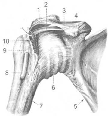
Fig. 58. Right shoulder joint (anterior view)
1 — acromion, 2 — coracohumeral ligament, 3 — coracoacromial ligament, 4 — coracoid process, 5 — scapula, 6 — joint capsule, 7 — humerus, 8 — tendon of the biceps brachii (long head), 9 — intertubercular synovial sheath, 10 — subscapularis muscle
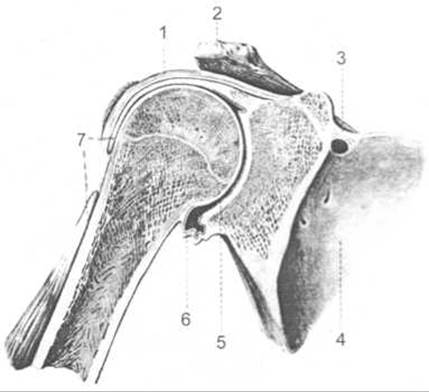
Fig. 59. Right shoulder joint (frontal section)
1 — joint capsule, 2 — acromion, 3 — superior transverse scapular ligament, 4 — scapula, 5 — joint cavity, 6 — joint capsule, 7 — tendon of the biceps brachii (long head)
The shoulder joint is reinforced by the coracohumeral and glenohumeral ligaments.
The coracohumeral ligament (lig. coracohumerale) is a thickening of the superior wall of the shoulder joint capsule that extends from the Base of the coracoid process of the scapula to the greater and lesser tubercles of the humerus.
The glenohumeral ligaments (ligg. glenohumeralia) appear as superior, middle, and inferior thickenings of the anterior wall of the joint capsule.
In addition, muscle fibers running close to the shoulder joint blend into its capsule. These include the supraspinatus, infraspinatus, subscapularis, and teres minor Muscles. These muscles not only reinforce the shoulder joint but also pull the relevant PARTS OF THE capsule away during movement, protecting it from pinching. Due to its high mobility and relatively moderate reinforcement, the shoulder joint is prone to frequent dislocations, including chronic ones.
The synovial layer of the shoulder joint capsule forms two constant outpouchings: the intertubercular synovial sheath and the subscapular bursa of the subscapularis muscle. The intertubercular synovial sheath (Vagina synovialis intertubercularis) is a finger-like outpocketing that surrounds the tendon of the long head of the biceps brachii as it passes across the joint over the head of the humerus. The subscapular bursa of the subscapularis muscle (bursa subtendinea musculi subscapularis) is located near the base of the coracoid process of the scapula, beneath the tendon of the subscapularis muscle. Normally, these outpouchings of the joint capsule are filled with synovial fluid, which reduces friction between the moving anatomical structures of the joint region.
The elbow joint (articulatio cubiti) is formed by the articular surfaces of the humerus, ulna, and radius (Figs. 60, 61). It is a compound joint comprising three individual joints of different shapes that share a common capsule and joint cavity: the humeroulnar, humeroradial, and proximal radioulnar joints.
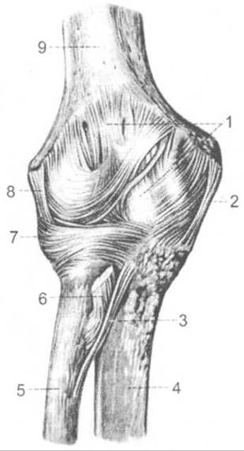
Fig. 60. Right elbow joint (anterior view)
1 — joint capsule, 2 — ulnar collateral ligament, 3 — oblique cord, 4 — ulna, 5 — radius, 6 — tendon of the biceps brachii (cut), 7 — annular ligament of the radius, 8 — radial collateral ligament, 9 — humerus.
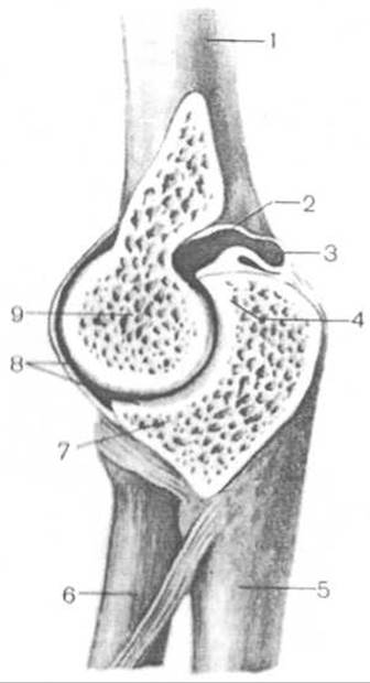
Fig. 61. Elbow joint (sagittal section)
1 — humerus, 2 — joint cavity, 3 — joint capsule, 4 — olecranon, 5 — ulna, 6 — radius, 7 — coronoid process, 8 — articular Cartilage, 9 — trochlea of the humerus.
The humeroulnar joint (articulatio humeroulnaris) is hinge-shaped, featuring a screw-like configuration of its articular surfaces. The humeral articular surface is formed by the trochlea of the humeral condyle. The groove (guidance groove) located on the trochlea is not perpendicular to the axis of the trochlea, but rather forms a certain angle with it. The trochlea articulates with the trochlear notch of the ulna, which bears a ridge corresponding to the groove on the humeral trochlea; consequently, movements around the frontal axis acquire a slightly spiral (screw-like) trajectory. The total range of motion (flexion and extension) reaches approximately 140°.
The humeroradial joint (articulatio humeroradialis) is formed by the small capitulum of the humeral condyle and the articular fovea of the head of the radius. Because it is ball-and-socket in shape, it is theoretically a triaxial joint. Movement around the frontal axis involves flexion and extension, occurring simultaneously with the same movements in the humeroulnar joint. Rotation — pronation and supination — occurs around the vertical axis simultaneously with identical movements in the proximal radioulnar joint. As for abduction and adduction around the sagittal axis, they are barely noticeable because the radius is bound to the ulna by the interosseous membrane and strong ligaments, preventing it from deviating away, while the ulna cannot follow it due to its constraint to the humerus via the hinge joint. Minimal movements around the sagittal axis do occur to the extent permitted by ligamentous elasticity. During maximal flexion, a slight abduction of the radius takes place, whereas during maximal extension, a slight adduction occurs.
The proximal radioulnar joint (articulatio radioulnaris proximalis) is formed by the articular circumference of the head of the radius and the radial notch of the ulna. This joint is cylindrical and combined in function (as it operates simultaneously with the distal radioulnar joint). It possesses a single vertical axis around which pronation and supination of the radius are possible within a range of 180°.
All the described joints are enclosed within a common articular capsule, which attaches: a) on the humerus anteriorly to the upper margin of the coronoid and radial fossae, posteriorly slightly below the upper margin of the olecranon fossa, and laterally between the condyle and epicondyles; b) on the radius to its neck; c) on the ulna to the margin of the trochlear notch. Anteriorly and posteriorly, the articular capsule is thin and lax.
The elbow joint is reinforced by the following ligaments:
— ulnar collateral ligament (lig. collaterale ulnare), which originates from the medial epicondyle of the humerus and attaches to the medial margin of the trochlear notch of the ulna;
— radial collateral ligament (lig. collaterale radii), which is thick and strong, originating from the lateral epicondyle of the humerus. Descending toward the head of the radius, it divides into two bundles: anterior and posterior. The anterior bundle runs anteriorly, while the posterior bundle runs posteriorly to the neck of the radius, encircling it in a loop-like manner. The anterior bundle attaches near the anterolateral margin of the trochlear notch of the ulna, whereas the posterior bundle blends into the annular ligament of the radius (lig. annulare radii). The latter consists of a bundle of arced fibrous fibers that encircle the neck of the radius and attach near the anterior and posterior margins of the radial notch of the ulna;
— quadrate ligament (lig. quadratum), which connects the distal margin of the radial notch of the ulna to the neck of the radius.
Last update: 08/08/2026
Editorial and Educational Adaptation: This material has been compiled based on the primary/original source text. The project team performed an editorial review, corrected technical inaccuracies, structured sections, and adapted the content for an educational format.
What was processed:
- elimination of formatting defects (OCR errors, structural breaks, corrupted characters);
- editorial organization of content;
- standardization of terminology in accordance with academic sources;
- verification of factual statements against the original source text.
All mentions of the author, publication year, and origin of the primary text have been preserved in accordance with the source.