Human Anatomy - Kotsan I. Y. 2009
Skeleton of the Upper Limb
Interosseous Joints of the Forearm
The proximal and distal ends of the radius and ulna of the forearm are connected by two joints: the proximal radioulnar and distal radioulnar joints. The space between the shafts of the forearm bones is bridged by the interosseous membrane of the forearm and the oblique cord (Fig. 62).
Class="center">
Fig. 62. Connections of the BONES OF THE (right) forearm, anterior view
1 — articular capsule, 2 — trochlea of the humerus, 3 — joint cavity, 4 — ulna, 5 — interosseous membrane of the forearm, 6 — distal radioulnar joint, 7 — radius, 8 — oblique cord, 9 — tendon of the biceps brachii Muscle (cut), 10 — annular ligament of the radius, 11 — HEAD of the radius, 12 — capitulum of the humerus, 13 — humerus
The interosseous membrane of the forearm (membrana interossea antebrachii) is formed by strong fibrous bundles that run obliquely downwards from the radius to the ulna. In the proximal and distal PARTS OF THE interosseous membrane, there are apertures for the passage of nerves and vessels. The interosseous membrane firmly connects the forearm bones to each other while not impeding the movement of the radius relative to the ulna. During supination of the forearm, the membrane becomes taut, whereas during pronation, it relaxes. A number of forearm Muscles originate from its PALMAR AND DORSAL surfaces.
The oblique cord (chorda obliqua) connects the ulnar tuberosity to the interosseous border of the radius and runs perpendicularly to the fibers of the interosseous membrane.
The distal radioulnar joint (articulatio radioulnaris distalis) is formed by the articular circumference of the ulnar head and the ulnar notch of the radius. Between the ulnar notch of the radius and the ulnar styloid process lies the articular disc (discus articularis) in the form of a triangular fibrocartilaginous plate. It separates the distal radioulnar joint from the radiocarpal joint and acts as a specialized articular socket for the head of the ulna. The articular capsule of the distal radioulnar joint is roomy, attaching along the margins of the articular surfaces and the articular disc, and forming a proximal pouch-like extension known as the sacciform recess (recessus sacciformis).
Together, the proximal and distal radioulnar joints form a combined, cylindrical-shaped joint. Movements in these joints occur around a longitudinal axis passing through the heads of the radius and ulna. In these joints, the radius rotates around the ulna. The maximum range of motion (supination and pronation) in the radioulnar joints reaches 180° (on average 140°).
The radiocarpal joint (articulatio radiocarpalis) is formed by the carpal articular surface of the radius, the distal surface of the articular disc completing the ulna, and the convex proximal articular surfaces of the three proximal Carpal Bones (scaphoid, lunate, and triquetrum) (Figs. 63, 64). The ulna does not take direct part in forming the radiocarpal joint, but only through the medium of Cartilage. Therefore, the joint is called radiocarpal rather than ulnocarpal. The radiocarpal joint is a compound, complex joint. Ellipsoid in shape, it has two axes of rotation: frontal and sagittal.
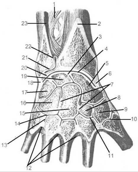
Fig. 63. Joints and ligaments of the hand (frontal section parallel to the dorsal surface of the hand; joint cavities opened)
1 — interosseous membrane of the forearm, 2 — radius, 3 — intercarpal ligament, 4 — radiocarpal joint, 5 — scaphoid bone, 6 — radial collateral ligament of the carpus, 7 — intercarpal ligaments, 8 — trapezoid bone, 9 — trapezium bone, 10 — carpometacarpal joint of the thumb, 11 — carpometacarpal joint, 12 — interosseous metacarpal ligaments, 13 — intermetacarpal joint, 14 — carpometacarpal joint, 15 — capitate bone, 16 — hamate bone, 17 — triquetral bone, 18 — ulnar collateral ligament, 19 — lunate bone, 20 — articular disc, 21 — radioulnar joint, 22 — sacciform recess, 23 — ulna
Flexion and extension of the hand are possible around the frontal axis, whereas adduction and abduction occur around the sagittal axis. Pronation and supination of the hand occur simultaneously with the corresponding Movements of the forearm bones. A slight passive rotational movement is also possible in the radioulnar joint due to the elasticity of the cartilage (10–12°). In addition, a minor circular movement (circumduction) is possible in the radiocarpal joint.

Fig. 64. Radiocarpal joint, ligaments, and JOINTS OF THE hand (right)
A — palmar surface: 1 — ulna, 2 — distal radioulnar joint, 3 — ulnar collateral ligament, 4 — pisiform bone, 5 — pisohamate ligament, 6 — pisometacarpal ligament, 7 — hook of the hamate bone, 8 — hamatometacarpal ligament, 9 — palmar carpometacarpal ligament, 10 — palmar metacarpal ligaments, 11 — deep transverse metacarpal ligaments, 12 — metacarpophalangeal joint (opened), 13 — fibrous Sheath of the digit, 14 — interphalangeal joints (opened), 15 — tendon of the flexor digitorum profundus muscle, 16 — tendon of the flexor digitorum superficialis muscle, 17 — fibrous sheath of the digit, 18 — collateral ligaments, 19 — carpometacarpal joint of the thumb, 20 — capitate bone, 21 — radiate carpal ligament, 22 — radial collateral ligament of the carpus, 23 — palmar radiocarpal ligament, 24 — lunate bone, 25 — radius, 26 — interosseous membrane of the forearm; B — dorsal surface: 1 — radial collateral ligament of the carpus, 2 — scaphoid bone, 3 — trapezium bone, 4 — carpometacarpal joint of the thumb, 5 — trapezoid bone, 6 — capitate bone, 7 — collateral ligaments, 8 — interphalangeal joint (opened), 9 — metacarpophalangeal joint, 10 — dorsal metacarpal ligaments, 11 — dorsal carpometacarpal ligaments, 12 — hamate bone, 13 — triquetral bone, 14 — ulnar collateral ligament of the carpus, 15 — dorsal radiocarpal ligament
The articular capsule of the radiocarpal joint is thin and attaches to the margins of the articular surfaces. On the radial side, the articular capsule is reinforced by the radial collateral ligament of the carpus (lig. collaterale carpi radiale), which extends from the radial styloid process to the scaphoid. On the ulnar side, the capsule is reinforced by the ulnar collateral ligament of the carpus (lig. collaterale carpi ulnare), stretching from the ulnar styloid process to the triquetrum and pisiform. On the palmar and dorsal surfaces of the radiocarpal joint lie the palmar and dorsal radiocarpal ligaments, respectively. The palmar radiocarpal ligament (lig. radiocarpeum palmare) originates from the anterior margin of the carpal articular surface of the radius and the base of its styloid process, runs downwards and medially, and attaches via separate bundles to the proximal carpal bones and the capitate bone of the distal row. The dorsal radiocarpal ligament (lig. radiocarpeum dorsale) extends from the dorsal surface of the distal end of the radius to the dorsum of the scaphoid, lunate, and triquetral bones. In addition to these ligaments, there are intercarpal interosseous ligaments (ligg. intercarpea interossea) connecting the bones of the proximal carpal row.
The midcarpal joint (articulatio mediocarpalis) is formed by the opposing articular surfaces of the proximal and distal rows of carpal bones and is functionally connected to the radiocarpal joint. The pisiform bone does not participate in forming this joint, as it forms an independent joint. The articular surfaces of this joint have a complex configuration, and the joint space on a frontal section of the hand has an S-shaped appearance. The cavity of the midcarpal joint extends between the bones forming the proximal and distal carpal rows, meaning it communicates with the cavities of the intercarpal joints. The midcarpal joint has limited mobility; its movement is closely linked with movement in the radiocarpal joint and allows only slight flexion and extension during maximal flexion and extension in the radiocarpal joint.
The intercarpal joints (articulationes intercarpales) are located between the individual carpal bones. They are formed by the opposing, articulating surfaces of the carpal bones.
The capsule of the intercarpal and midcarpal joints is thin and is reinforced by the following ligaments.
1. Radiate carpal ligament (lig. carpi radiatum). It consists of a collection of fiber bundles originating from the capitate bone and radiating to the adjacent bones of the proximal and distal carpal rows.
2. Palmar intercarpal ligaments (ligg. intercarpalia palmaria) — short ligaments stretched between the carpal bones on the palmar side.
3. Dorsal intercarpal ligaments (ligg. intercarpalia dorsalia) stretched between individual carpal bones on the dorsal side.
The individual carpal bones are connected by intracapsular ligaments, namely the intercarpal interosseous ligaments (ligg. intercarpalia interossea).
The pisiform joint (articulatio ossis pisiformis) connects the pisiform bone to the triquetrum. The sesamoid pisiform bone possesses an articular surface only on the side that contacts the opposing articular surface of the triquetrum. The articular capsule is fixed to the articular surfaces of the bones. The joint cavity may communicate with the cavity of the radiocarpal joint. The joint is reinforced by the pisohamate ligament (lig. pisohamatum), running from the pisiform bone to the hook of the hamate bone, and the pisometacarpal ligament (lig. pisometacarpale), running from the pisiform bone to the bases of the third to fifth Metacarpal bones. Both ligaments are continuations of the tendon of the flexor carpi ulnaris muscle, within the substance of which the pisiform bone itself is embedded.
The carpometacarpal joints (articulationes carpometacarpales) connect the bones of the distal carpal row with the bases of the metacarpal bones. There are two structurally and functionally distinct carpometacarpal joints: the carpometacarpal joint of the thumb and the carpometacarpal joints of the second through fifth metacarpal bones.
The carpometacarpal joint of the thumb (articulatio carpometacarpalis pollicis) is formed by the distal articular surface of the trapezium bone and the articular surface of the Base of the first metacarpal bone. It is a simple, saddle-shaped joint featuring two mutually perpendicular axes of rotation: the frontal and the sagittal axes. Flexion and extension of the thumb, along with its metacarpal bone, occur around the frontal axis; however, because this axis is not entirely transverse, the thumb shifts toward the palm during flexion, opposing the little finger and other digits. This movement is referred to as opposition (oppositio), while the reverse movement is termed reposition (reppositio). Abduction and adduction of the thumb toward the index finger take place around the sagittal axis. As a result of combining these movements around the frontal and sagittal axes, circumduction becomes possible within the joint.
The carpometacarpal joints of the II–V metacarpal bones (articulationes carpometacarpeae) are formed by the articular surfaces of the distal carpal row and the bases of the II–V metacarpal bones. These are plane joints whose shared articular cavity is narrow and stepped at right angles. It branches into the spaces between the bases of the metacarpal bones (intermetacarpal joints) and between the carpal bones (intercarpal joints), and also communicates with the cavity of the midcarpal joint. These joints have very limited mobility, allowing only slight gliding movements.
The intermetacarpal joints (articulationes intermetacarpales) are formed by the adjacent surfaces of the bases of the II–V metacarpal bones. They are simple, plane, multiaxial joints with limited mobility. The joint cavity communicates with the cavity of the carpometacarpal joints. The ligaments reinforcing these joints are stretched transversely between the bases of the metacarpal bones and include the following:
1. Palmar metacarpal ligaments (ligg. metacarpalia palmaria), three in number, connecting the bases of the II–V metacarpal bones.
2. Dorsal metacarpal ligaments (ligg. metacarpalia dorsalia), four in number, connecting the bases of all metacarpal bones.
3. Interosseous metacarpal ligaments (ligg. metacarpalia interossea) — four intracapsular ligaments connecting the bases of the I–V metacarpal bones.
The metacarpophalangeal joints (articulationes metacarpophalangeales) are formed by the articular surfaces of the heads of the I–V metacarpal bones and the articular sockets of the bases of the proximal Phalanges of the I–V digits. The first metacarpophalangeal joint (of the thumb) is classified as an ellipsoid joint, allowing movement around two axes: frontal and sagittal. The metacarpophalangeal joints of the second through fifth digits are spheroidal (ball-and-socket) in shape, permitting movement around three axes. Flexion and extension of the digits occur around the frontal axis, whereas abduction (away from the middle digit) and adduction take place around the sagittal axis. Pronation and supination around the vertical axis can occur only passively under the Influence of External forces. Active movements (pronation and supination) are impossible due to the absence of dedicated muscles, as well as the presence of ligaments that restrict these movements. Circumduction is also possible in these joints.
The articular capsules are spacious and reinforced laterally by collateral ligaments (ligg. collateralia). On the palmar side, the capsules are thickened by bundles of fibers from the palmar ligaments (ligg. palmaria). Additionally, the metacarpophalangeal joints of the II–V digits are reinforced by deep transverse metacarpal ligaments (ligg. metacarpea transversa profunda), the fibers of which span between the heads of the II–V metacarpal bones.
The interphalangeal joints of the hand (articulationes interphalangeales) are formed by the heads and bases of adjacent phalanges. All these joints have a similar Structure and, based on the shape of their articular surfaces, are typical hinge (ginglymus) joints. Movements in these joints occur exclusively around the frontal axis in the form of flexion and extension.
The capsule of each joint is loose; it is reinforced laterally by collateral ligaments (ligg. collateralia) and on the palmar side by palmar ligaments (ligg. palmaria).
Basic Latin Terms
Pectoral Girdle — cingulum membri superioris
Free part of the upper limb — pars libera membri superioris
Scapula — scapula
Costal (anterior) surface — facies costalis (anterior)
Subscapular fossa — fossa subscapularis
Posterior surface — facies posterior
Spine of scapula — spina scapulae
Supraspinous fossa — fossa supraspinata
Infraspinous fossa — fossa infraspinata
Acromion — acromion
Articular facet of acromion — facies articularis acromii
Medial border — margo medialis
Lateral border — margo lateralis
Superior border — margo superior
Scapular notch — incisura scapulae
Inferior angle — angulus inferior
Lateral angle — angulus lateralis
Superior angle — angulus superior
Glenoid cavity — cavitas glenoidalis
Supraglenoid tubercle — tuberculum supraglenoidale
Infraglenoid tubercle — tuberculum infraglenoidale
Neck of scapula — collum scapulae
Coracoid process — processus coracoideus
Clavicle — clavicula
Sternal end — extremitas sternalis
Sternal articular surface — facies articularis sternalis
Impression for costoclavicular ligament — impressio ligamentum costoclavicularis
Body of clavicle — corpus claviculae
Acromial end — extremitas acromialis
Articular surface of acromion — facies articularis acromialis
Conoid tubercle — tuberculum conoideum
Trapezoid line — linea trapezoidea
Humerus — humerus
Head of humerus — caput humeri
Anatomical neck — collum anatomicum
Surgical neck — collum chirurgicum
Greater tubercle — tuberculum majus
Lesser tubercle — tuberculum minus
Intertubercular sulcus — sulcus intertubercularis
Crest of greater tubercle — crista tuberculi majoris
Crest of lesser tubercle — crista tuberculi minoris
Body of humerus — corpus humeri
Anteromedial surface — facies anterior medialis
Anterolateral surface — facies anterior lateralis
Posterior surface — facies posterior
Radial sulcus — sulcus nervus radialis
Medial border — margo medialis
Medial supracondylar ridge — crista supracondylaris medialis
Lateral border — margo lateralis
Lateral supracondylar ridge — crista supracondylaris lateralis
Deltoid tuberosity — tuberositas deltoidea
Condyle of humerus — condylus humeri
Capitulum of humerus — capitulum humeri
Trochlea of humerus — trochlea humeri
Olecranon fossa — fossa olecrani
Coronoid fossa — fossa coronoidea
Radial fossa — fossa radialis
Medial epicondyle — epicondylus medialis
Ulnar nerve sulcus — sulcus nervus ulnaris
Lateral epicondyle — epicondylus lateralis
Radius — radius
Head of radius — caput radii
Articular facet — fovea articularis
Articular circumference — circumferentia articularis
Neck of radius — collum radii
Body of radius — corpus radii
Radial tuberosity — tuberositas radii
Anterior surface — facies anterior
Posterior surface — facies posterior
Lateral surface — facies lateralis
Interosseous border — margo interosseus
Anterior border — margo anterior
Posterior border — margo posterior
Styloid process — processus styloideus
Dorsal tubercle — tuberculum dorsale
Ulnar notch — incisura ulnaris
Carpal articular surface — facies articularis carpalis
Ulna — ulna
Olecranon — olecranon
Coronoid process — processus coronoideus
Trochlear notch — incisura trochlearis
Ulnar tuberosity — tuberositas ulnae
Radial notch — incisura radialis
Body of ulna — corpus ulnae
Anterior surface — facies anterior
Posterior surface — facies posterior
Medial surface — facies medialis
Interosseous border — margo interosseus
Anterior border — margo anterior
Posterior border — margo posterior
Supinator crest — crista musculi supinatoris
Head of ulna — caput ulnae
Articular circumference — circumferentia articularis
Styloid process — processus styloideus
Bones of the hand — ossa manus
Carpal bones — carpus
Scaphoid bone — os scaphoideum
Tubercle of scaphoid — tuberculum ossis scaphoidei
Lunate bone — os lunatum
Triquetral bone — os triquetrum
Pisiform bone — os pisiforme
Trapezium bone — os trapezium
Tubercle of trapezium — tuberculum ossis trapezii
Trapezoid bone — os trapezoideum
Capitate bone — os capitatum
Hamate bone — os hamatum
Hook of hamate — hamulus ossis hamati
Carpal sulcus — sulcus carpi
Metacarpus — metacarpus
Base of metacarpal bone — basis metacarpalis
Body of metacarpal bone — corpus metacarpale
Head of metacarpal bone — caput metacarpale
Bones of the digits (phalanges) — ossa digitorum (phalanges)
Proximal phalanx — phalanx proximalis
Middle phalanx — phalanx media
Distal phalanx — phalanx distalis
Base of phalanx — basis phalangis
Body of phalanx — corpus phalangis
Head of phalanx — caput phalangis
Sternoclavicular joint — articulatio sternoclavicularis
Anterior sternoclavicular ligament — lig. sternoclaviculare anterius
Posterior sternoclavicular ligament — lig. sternoclaviculare posterius
Costoclavicular ligament — lig. costoclaviculare
Interclavicular ligament — lig. interclaviculare
Acromioclavicular joint — articulatio acromioclavicularis
Acromioclavicular ligament — lig. acromioclaviculare
Coracoclavicular ligament — lig. coracoclaviculare
Trapezoid ligament — lig. trapezoideum
Conoid ligament — lig. conoideum
Coracoacromial ligament — lig. coracoacromiale
Superior transverse scapular ligament — lig. transversum scapulae superius
Inferior transverse scapular ligament — lig. transversum scapulae inferius
Shoulder joint — articulatio humeri
Glenoid labrum — labrum glenoidale
Glenohumeral ligaments — ligg. glenohumeralia
Coracohumeral ligament — lig. coracohumerale
Intertubercular synovial sheath — Vagina synovialis intertubercularis
Subtendinous bursa of subscapularis — bursa subtendinea
subscapularis muscle
Elbow joint — articulatio cubiti
Humeroulnar joint — articulatio humeroulnaris
Humeroradial joint — articulatio humeroradialis
Proximal radioulnar joint — articulatio radioulnaris proximalis
Ulnar collateral ligament — lig. collaterale ulnare
Radial collateral ligament — lig. collaterale radii
Anular ligament of radius — lig. annulare radii
Interosseous membrane of forearm — membrana interossea antebrachii
Oblique cord — chorda obliqua
Distal radioulnar joint — articulatio radioulnaris distalis
Sacciform recess — recessus sacciformis
Radiocarpal joint — articulatio radiocarpalis
Dorsal radiocarpal ligament — lig. radiocarpale dorsale
Palmar radiocarpal ligament — lig. radiocarpale palmare
Palmar ulnocarpal ligament — lig. ulnocarpale palmare
Ulnar collateral ligament of wrist — lig. collaterale carpi ulnare
Radial collateral ligament of wrist — lig. collaterale carpi radiale
Midcarpal joint — articulatio mediocarpalis
Intercarpal joints — articulationes intercarpales
Radiate carpal ligament — lig. carpi radiatum
Dorsal intercarpal ligaments — ligg. intercarpalia dorsalia
Palmar intercarpal ligaments — ligg. intercarpalia palmaria
Interosseous intercarpal ligaments — ligg. intercarpalia interossea
Joint of pisiform bone — articulatio ossis pisiformis
Pisohamate ligament — lig. pisohamatum
Pisometacarpal ligament — lig. pisometacarpale
Carpometacarpal joints — articulationes carpometacarpales
Dorsal carpometacarpal ligaments — ligg. carpometacarpalia dorsalia
Palmar carpometacarpal ligaments — ligg. carpometacarpalia palmaria
Carpometacarpal joint of the thumb — articulatio carpometacarpalis pollicis
Intermetacarpal joints — articulationes intermetacarpales
Dorsal metacarpal ligaments — ligg. metacarpalia dorsalia
Palmar metacarpal ligaments — ligg. metacarpalia palmaria
Interosseous metacarpal ligaments — ligg. metacarpalia interossea
Metacarpophalangeal joints — articulationes metacarpophalangeales
Collateral ligaments — ligg. collateralia
Palmar ligaments — ligg. palmaria
Deep transverse metacarpal ligament — lig. metacarpale transversum profundum
Interphalangeal joints of the hand — articulationes interphalangeales manus
Collateral ligaments — ligg. collateralia
Palmar ligaments — ligg. palmaria
Last update: 08/08/2026
Editorial and Educational Adaptation: This material has been compiled based on the primary/original source text. The project team performed an editorial review, corrected technical inaccuracies, structured sections, and adapted the content for an educational format.
What was processed:
- elimination of formatting defects (OCR errors, structural breaks, corrupted characters);
- editorial organization of content;
- standardization of terminology in accordance with academic sources;
- verification of factual statements against the original source text.
All mentions of the author, publication year, and origin of the primary text have been preserved in accordance with the source.