Human Anatomy - Kotsan I. Ya. 2009
Skeleton of the Upper Limb
Joints of the Bones of the Pectoral Girdle
The BONES OF THE Pectoral Girdle are connected by two joints: the sternoclavicular and acromioclavicular joints.
Class="center">
Fig. 56. Sternoclavicular joints
(anterior view, the right sternoclavicular joint is shown in a frontal section)
1 — articular disc, 2 — interclavicular ligament, 3 — sternoclavicular ligament, 4 — clavicle, 5 — first rib, 6 — costoclavicular ligament, 7 — manubrium of Sternum
The sternoclavicular joint (articulatio sternoclavicularis) is formed by the articular surface of the sternal end of the clavicle and the clavicular notch of the manubrium of the sternum (Fig. 56). The articular surfaces of this connection have low congruence. The joint is simple, complex, and bicompartmental (due to the presence of an articular disc in the joint cavity, which divides it into two compartments). The shape of the joint approximates a saddle joint; however, thanks to the articular disc—which smooths out inequalities and helps increase the congruence of the articular surfaces—movements in this joint can occur around three axes: the sagittal axis (elevation and depression of the clavicle), the vertical axis (forward and backward movement of the clavicle), and the frontal axis (slight Rotation of the clavicle around its own longitudinal axis). In addition, circular movement (circumduction) is possible in this joint, during which the acromial end of the clavicle describes an ellipse.
Of all the bones of the pectoral girdle, only the clavicle is connected to the Axial Skeleton. Therefore, when the clavicle moves, the scapula connected to it and the entire free upper limb move along with it.
The capsule of the sternoclavicular joint is thin and attaches along the margins of the articular surfaces.
The joint is reinforced by the following ligaments:
— the anterior sternoclavicular ligament (lig. sternoclaviculare anterius), which is a thickening of the anterior wall of the Joint Capsule;
— the posterior sternoclavicular ligament (lig. sternoclaviculare posterius), which is a thickening of the posterior wall of the joint capsule;
— the interclavicular ligament (lig. interclaviculare), which connects both clavicles, filling the jugular notch;
— the costoclavicular ligament (lig. costoclaviculare), which extends from the Cartilage of the first rib to the Inferior surface of the clavicle and checks upward Displacement of the clavicle. In some individuals (most frequently in those engaged in heavy physical labor), a costoclavicular joint may form at the site of the strong costoclavicular ligament.
The acromioclavicular joint (articulatio acromioclavicularis) is formed by the acromial articular surface of the clavicle and the articular surface of the acromion of the scapula (Fig. 57). Both articular surfaces are slightly curved. Frequently, an articular disc is present within the joint cavity, dividing it into two compartments. The articular disc is composed of fibrocartilage, attaches to the fibrous layer of the joint capsule, and may contain an aperture. The joint is simple and plane in shape. Movements in the joint occur around three axes, but their range is minimal because the ligaments restrict joint mobility.
The acromioclavicular joint is reinforced by the acromioclavicular and coracoclavicular ligaments. The acromioclavicular ligament (lig. acromioclaviculare) is stretched between the acromial end of the clavicle and the acromial process of the scapula, reinforcing the joint capsule superiorly. The coracoclavicular ligament (lig. coracoclaviculare) consists of two parts: the trapezoid ligament (lig. trapezoideum) and the conoid ligament (lig. conoideum), which extend from the coracoid process of the scapula to the trapezoid line and conoid tubercle of the clavicle, respectively.
In addition to these ligaments, the scapula has its own proper ligaments that connect its individual parts but are not associated with joints. These include:
— the coracoacromial ligament (lig. coracoacromiale), which has the appearance of a triangular band stretched like an arch over the shoulder joint between the coracoid and acromial processes of the scapula. This ligament protects the shoulder joint from above and limits upward movement of the humerus during abduction of the arm;
— the superior transverse scapular ligament (lig. transversum scapulae superius), which bridges across the scapular notch and converts it into a foramen through which the suprascapular nerve passes;
— the inferior transverse scapular ligament (lig. transversum scapulae inferius), which connects the Base of the acromial process of the scapula with the posterior margin of the glenoid cavity of the scapula. The descending branch of the suprascapular artery passes beneath this ligament.
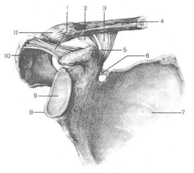
Fig. 57. Acromioclavicular joint (right) and ligaments of the scapula
1 — coracoacromial ligament, 2 — trapezoid ligament, 3 — conoid ligament, 4 — acromial end of the clavicle, 5 — coracoid process, 6 — superior transverse scapular ligament, 7 — scapula, 8 — glenoid labrum, 9 — glenoid cavity, 10 — acromion, 11 — acromioclavicular joint (acromioclavicular ligament visible).
Last update: 08/08/2026
Editorial and Educational Adaptation: This material has been compiled based on the primary/original source text. The project team performed an editorial review, corrected technical inaccuracies, structured sections, and adapted the content for an educational format.
What was processed:
- elimination of formatting defects (OCR errors, structural breaks, corrupted characters);
- editorial organization of content;
- standardization of terminology in accordance with academic sources;
- verification of factual statements against the original source text.
All mentions of the author, publication year, and origin of the primary text have been preserved in accordance with the source.