Human Anatomy and Physiology - N. I. Fedyukovich 2003
Bones and Joints
Skeleton of the Limbs
Bones of the Upper Limb
Pectoral Girdle. The scapula (shoulder blade) is a flat, triangular bone located on the posterior aspect of The thoracic cage at the level of the II–VIII Ribs (Figs. 36, 37)
Class="center">
Fig. 36. Right scapula (posterior view):
1 — coracoid process; 2 — acromion; 3 — lateral angle; 4 — neck of the scapula; 5 — lateral border; 6 — inferior angle; 7 — medial border; 8 — subscapular fossa; 9 — posterior surface; 10 — spine of the scapula; 11 — supraspinous fossa; 12 — scapular notch; 13 — superior angle
The scapula features costal and dorsal surfaces, superior, inferior, and lateral angles, as well as superior, lateral (outer), and medial (inner) borders. The dorsal (posterior) surface of the scapula is divided by the spine of the scapula into the supraspinous and infraspinous fossae; the spine of the scapula continues into the acromion. The scapula also has an articular surface (glenoid cavity) for articulation with the humerus and an anteriorly directed coracoid process.

Fig. 37. Right scapula (anterior view):
1 — articular surface of the acromion; 2 — scapular notch; 3 — spine of the scapula; 4 — superior angle; 5 — medial border; 6 — inferior angle; 7 — lateral border; 8 — subscapular fossa; 9 — costal surface; 10 — neck of the scapula; 11 — infraglenoid tubercle; 12 — lateral angle; 13 — medial border; 14 — supraglenoid tubercle; 15 — coracoid process
The clavicle (clavicula) is an S-shaped bone consisting of a body, as well as acromial and sternal ends with articular surfaces (Fig. 38).

Fig. 38. Right clavicle (inferior view):
1 — acromial articular surface; 2 — trapezoid line; 3 — subclavian groove; 4 — body of the clavicle; 5 — sternal end; 6 — sternal articular surface; 7 — impression for costoclavicular ligament; 8 — conoid tubercle; 9 — acromial end
The former articulates with the acromion of the scapula, and the latter with the Sternum.
JOINTS OF THE pectoral girdle. The sternal end of the clavicle connects to the sternum via the sternoclavicular joint (Fig. 39). Due to the presence of an intra-articular articular disc, movement in the joint occurs around the sagittal axis (upward and downward) and around the vertical axis (forward and backward). Thus, slight circumduction is possible. The acromial end of the clavicle articulates with the acromion, forming the acromioclavicular joint. It is a plane joint with a small range of motion, strongly reinforced by the Joint Capsule and ligaments—the acromioclavicular and coracoclavicular ligaments.

Fig. 39. Sternoclavicular joints:
1 - costoclavicular ligament; 2 - anterior sternoclavicular ligament; 3 - interclavicular ligament; 4 - articular disc.
Skeleton of the free upper limb. The humerus belongs to the long bones and has a shaft (body), as well as proximal and distal ends (Figs. 40, 41).
The thickened proximal end forms the HEAD of the humerus. Along the margin of the head runs a shallow groove—the anatomical neck, near which lie the greater and lesser tubercles, separated by a groove. The narrowest part between the head of the humerus and its shaft is called the surgical neck (a common site of fractures). The distal end of the humerus is expanded, forming the condyle of the humerus, flanked by two projections—the medial and lateral epicondyles. The medial part of the condyle forms the trochlea of the humerus for articulation with the ulna of the forearm. Lateral to the trochlea is the capitulum of the humerus, which articulates with the radius. Ligaments and Muscles attach to the greater and lesser tubercles, epicondyles, and other structures of the humerus.

Fig. 40. Right humerus (anterior view):
1 — head of the humerus; 2 — anatomical neck; 3 — anteromedial surface; 4 — medial border; 5 — condyle of the humerus; 6 — medial epicondyle; 7 — trochlea of the humerus; 8 — capitulum of the humerus; 9 — lateral epicondyle; 10 — lateral border; 11 — body of the humerus; 12 — deltoid tuberosity; 13 — anterolateral surface; 14 — surgical neck; 15 — lesser tubercle; 16 — greater tubercle

Fig. 41. Right humerus (posterior view):
1 — head of the humerus; 2 — anatomical neck; 3 — greater tubercle; 4 — surgical neck; 5 — condyle of the humerus; 6 — lateral epicondyle; 7 — olecranon fossa; 8 — trochlea of the humerus; 9 — groove for ulnar nerve; 10 — medial epicondyle
The BONES OF THE forearm consist of two long tubular bones—the radius and the ulna (Fig. 42). Each bone has a shaft (diaphysis) and two ends (epiphyses).
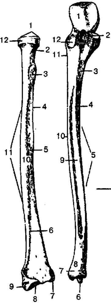
Fig. 42. Right radius and ulna (anterior view):
A — radius: 1 — head of radius; 2 — neck of radius; 3 — radial tuberosity; 4 — interosseous border; 5 — anterior surface; 6 — anterior border; 7 — ulnar notch; 8 — carpal articular surface; 9 — styloid process; 10 — lateral surface; 11 — shaft of radius; 12 — articular circumference; B — ulna: 1 — trochlear notch; 2 — coronoid process; 3 — ulnar tuberosity; 4 — anterior border; 5 — shaft of ulna; 6 — styloid process; 7 — articular circumference; 8 — head of ulna; 9 — anterior surface; 10 — interosseous border; 11 — supinator crest; 12 — radial notch
The radius is located on the lateral side of the forearm. Its upper end forms the head with an articular fovea and an articular circumference that articulates with the radial notch of the ulna. The lower end has a concave carpal articular surface for articulation with the proximal row of Carpal Bones. The shafts and epiphyses of the forearm bones feature bony prominences for the attachment of muscles and ligaments.
The ulna is located medially and has a trihedral shape, presenting anterior, posterior, and medial surfaces. Its thickened upper end features two notches—the radial and trochlear notches. The latter is bounded by the coronoid and olecranon processes and is designed to articulate with the trochlea of the humerus. The lower end of the ulna has a head, an articular circumference, and a styloid process.
The bones of the hand are divided into the carpal bones, Metacarpal bones, and Phalanges (bones of the digits) (Fig. 43).

Fig. 43. Bones of the right hand (dorsal surface):
1 — distal phalanx; 2 — middle phalanx; 3 — head of phalanx; 4 — phalanges (bones of the digits); 5 — proximal phalanx; 6 — base of phalanx; 7 — shaft of phalanx; 8 — head of metacarpal bone; 9 — third metacarpal bone; 10 — shaft of metacarpal bone; 11 — base of metacarpal bone; 12 — metacarpus (1—У metacarpal bones); 13 — styloid process; 14 — trapezium; 15 — trapezoid bone; 16 — capitate bone; 17 — hamate bone; 18 — triquetral bone; 19 — pisiform bone; 20 — lunate bone; 21 — scaphoid bone
The carpal bones (ossa carpi) consist of short cancellous bones arranged in two rows of four. The proximal row consists of the pisiform, triquetral, lunate, and scaphoid bones, while the distal row consists of the hamate, capitate, trapezoid, and trapezium bones. The palmar surface of the carpus is slightly concave, forming a groove covered by a ligament. This ligament converts the carpal groove into a canal through which Muscle tendons and nerves pass.
The metacarpal bones (ossa metacarpi) are five short tubular bones. Each consists of a base, a shaft, and a head. The base and head have articular surfaces for articulation with the carpal bones and the phalanges of the digits, respectively.
The bones of the digits (ossa digitorum) consist of short tubular bones called phalanges. Each digit, except for the thumb, has three phalanges: proximal, middle, and distal. The thumb has only two phalanges: proximal and distal.
Joints of the upper limb. The joints of the free upper limb connect its bones to each other and to the pectoral girdle.
The shoulder joint (articulatio humeri) is formed by the head of the humerus and the glenoid cavity of the scapula, which is deepened by the glenoid labrum (Fig. 44).

Fig. 44. Shoulder joint (longitudinal section):
1 — tendon of the long head of the biceps brachii muscle; 2, 7 — joint capsule; 3 — acromion of the scapula; 4 — superior transverse scapular ligament; 5 — scapula; 6 — glenoid cavity of the scapula
The joint capsule encloses the head of the humerus at the anatomical neck, and on the scapula, it attaches along the margin of the glenoid cavity. The joint is reinforced by the coracohumeral ligament and surrounding muscles. The tendon of the long head of the biceps brachii muscle passes through the joint cavity. The shoulder joint is a ball-and-socket joint, allowing movement around three axes: frontal, sagittal, and vertical.
The elbow joint (articulatio cubiti) is a compound joint comprising the humeroulnar, humeroradial, and proximal radioulnar joints. These three joints share a single joint capsule, which is reinforced by the radial and ulnar collateral ligaments, as well as the anular ligament of the radius. The elbow joint is classified as a hinge joint, allowing flexion, extension, and Rotation of the forearm (Fig. 45).
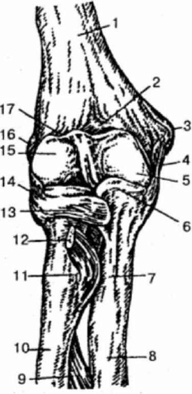
Fig. 45. Right elbow joint (anterior view):
1 — humerus; 2 — coronoid fossa; 3 — medial epicondyle; 4, 13, 14 — ligaments; 5 — trochlea of humerus; 6 — coronoid process; 7 — ulnar tuberosity; 8 — ulna; 9 — interosseous membrane of forearm; 10 — radius; 11 — radial tuberosity; 12 — tendon of biceps brachii muscle; 15 — capitulum of humerus; 16 — lateral epicondyle; 17 — radial fossa
The distal radioulnar joint (articulatio radioulnaris distalis) is an independent joint, whereas the proximal radioulnar joint is part of the elbow joint. Together, however, they form a single combined pivot (trochoid) joint. If the rotation of the radius around its longitudinal axis, along with the palmar surface of the hand, is directed medially, this movement is called pronation; the opposite movement is supination.
The wrist joint (articulatio radiocarpalis) is a compound ellipsoid joint formed by the carpal articular surface of the radius and three bones of the proximal row of the carpus. It allows two MAIN TYPES OF movement: adduction and abduction, flexion and extension, as well as slight passive circumduction. The joint is enclosed by a common capsule and reinforced by strong ulnar, radial, palmar, and dorsal radiocarpal ligaments.
The joints of the hand include the intermetacarpal, carpometacarpal, metacarpophalangeal, and interphalangeal joints. These joints are reinforced by short interosseous ligaments located on the PALMAR AND DORSAL surfaces of the hand, outside the joint cavities. The carpometacarpal joint of the thumb has a unique Structure. It is saddle-shaped and permits two axes of movement: flexion and extension, adduction and abduction; circumduction is also possible, as well as opposition of the thumb to the other fingers. The metacarpophalangeal joints are condyloid, while the interphalangeal joints are hinge joints. The Structural Features of the Bones and joints of the hand account for its extraordinary mobility, allowing it to perform highly precise and diverse movements.
Last update: 08/08/2026
Editorial and Educational Adaptation: This material has been compiled based on the primary/original source text. The project team performed an editorial review, corrected technical inaccuracies, structured sections, and adapted the content for an educational format.
What was processed:
- elimination of formatting defects (OCR errors, structural breaks, corrupted characters);
- editorial organization of content;
- standardization of terminology in accordance with academic sources;
- verification of factual statements against the original source text.
All mentions of the author, publication year, and origin of the primary text have been preserved in accordance with the source.