Human Anatomy, Part 1 - K. A. Dyubenko, A. K. Kolomiytsev, Yu. B. Chaikovsky 2002
Special Part
Splanchnology, splanchnologia [the study of viscera] — Digestive system, systema digestorium
Intestine — Small intestine — Jejunum and ileum
The jejunum and ileum are located in the middle and lower Abdominal cavity, completely covered by the Peritoneum intraperitoneally, and possess a mesentery, mesenterium. The initial part of the jejunum has limited mobility due to its short mesentery and the fixation of the duodenojejunal flexure. The loops of the jejunum lie horizontally in the upper left part of the lower abdominal floor and in the umbilical region, regio umbilicalis. The loops of the ileum are positioned vertically in the hypogastric region, regio hypogastńcum, the right iliac fossa, and the pelvic cavity. The distal segment of the ileum empties into the cecum. Approximately 1 m from its termination, a 5-7 cm long appendage may be present—the Meckel's diverticulum, diverticulum ilei (Meckeli)—a congenital anomaly occurring in 2% of individuals (Richard M. Mendelson, 1996) that can lead to acute inflammation, melena, and intestinal volvulus. Anteriorly, the loops of the Small Intestine are covered by the greater omentum; to the right by the ascending colon; superiorly by the transverse colon; and to the left by the descending colon. Posteriorly, the jejunal and ileal loops lie against the parietal peritoneum. Inferiorly and to the left, the small intestine loops are adjacent to the sigmoid colon, and in the pelvic cavity, they relate to the Urinary Bladder and rectum in men, and to the Uterus in women. The jejunum can be distinguished from the ileum by its larger diameter (4-6 cm compared to 3-3.5 cm in the ileum). The wall of the jejunum is thicker, has a redder color, and its mucous membrane forms a greater number of folds and villi.
Structure OF THE small intestine. The wall of the small intestine consists of four tunics (Fig. 232):
- mucous membrane, tunica mucosa;
- submucosa, tela submucosa;
- muscular coat, tunica mиslularis;
- serous coat, tunica serosa.
The mucous membrane, tunica mucosa, is formed by a single-layered cylindrical and prismatic epithelium, the submucosa, and the muscular layer of the mucosa. The relief of the mucous membrane has a characteristic velvety appearance due to the presence of circular folds, intestinal villi, and crypts. The small intestine contains about 650-700 folds, reaching up to 5 cm in length and 8 mm in height. In the duodenum, besides circular folds, There is a longitudinal fold, plica longitudinalis duodeni, which originates on the left wall of the descending part and ends at the major duodenal papilla (Vater), papilla duodeni major (Voten). The common Bile duct and pancreatic duct open into this papilla via a single common orifice. This anatomical landmark is of clinical importance in surgical practice.
Class="center">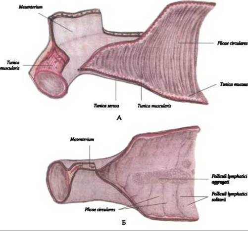
Fig. 232. Small intestine (according to R. D. Sinelnikov):
A - mucous membrane, tunica mucosa of the jejunum; B - mucous membrane, tunica mucosa of the ileum
Intestinal villi, villi intestinales (Fig. 233), are finger-like projections of the mucous membrane lacking a submucosal layer. There are about 4-5 million of them in the small intestine. Their primary function is the absorption of Proteins, fats, and CARBOHYDRATES, as well as increasing the absorptive and secretory surface area of the small intestine. In the duodenum and jejunum, their number can reach 30-40 per 1 mm2. Each villus contains Blood and Lymphatic vessels that form vascular networks, as well as nerve fibers. The activity of the villi is regulated by the hormone villikinin.
The surface of the villi is covered by a single-layered columnar epithelium, which comprises three Cell types: brush-border enterocytes, mucus-producing goblet Cells, and a small number of enteroendocrine cells.

Fig. 233. Intestinal villi, villi intestinalis (according to F. Kiss and J. Szentágothai)
The Role of microvilli is not limited to this—they contain A large number of active Enzymes involved in membrane Digestion (contact digestion) and the absorption of nutrients. Intestinal crypts, or glands, cryptae intestinales seu glandulae Lieberkuchni, are tubular invaginations of the epithelium extending into the lamina propria of the mucous membrane (Fig. 234). Their length reaches 0.5 mm, with up to 100 crypts per 1 mm2. They are most abundant in the duodenum and jejunum, and less numerous in the ileum. The total surface area of the crypts in the small intestine is 14 m2. Food entering the small intestine undergoes digestive processes not only within the intestinal lumen but also between the microvilli and within the crypts. In the submucosa, aggregations of lymphoid tissue form solitary lymphatic nodules, noduli lymphaidei solitarii, and aggregated lymphatic follicles [Peyer's patches], lymphoidei noduli aggregati (Peyeri).

Fig. 234. Intestinal crypt, cryptae intestinalis (according to F. Peter)
Solitary follicles are distributed evenly throughout the entire length of the small intestine, ranging in diameter from 0.5 to 3 mm. Aggregated follicles have a length of 2 to 12 cm, a width of 1-3 cm, and are located in the mucous membrane of the ileum opposite the mesenteric attachment. The total number of Aggregated lymphoid follicles ranges from 20 to 30.
The submucosa, tela submucosa, of the duodenum and the initial part of the jejunum contains numerous branched tubular duodenal glands, glandulae duodenalis, which produce intestinal juice. Additionally, the small intestine contains a large number of simple tubular intestinal glands, glandulae intestinales, which secrete intestinal juice and mucus. Furthermore, Major Digestive Glands—The Liver and Pancreas—play a crucial role in The production of intestinal juice.
The muscular coat, tunica muscularis, consists of longitudinal and circular smooth Muscle fibers, with the circular layer being the most developed. Between the layers lies a stratum of unformed Connective Tissue containing vascular networks and a nerve plexus. The main function of the muscular coat is to mix the chyme within the intestinal lumen and propel it in a distal direction. This is achieved through the coordinated action of the longitudinal and circular layers of the intestinal wall. The bundles of smooth muscle fibers in both layers are spirally oriented and perform Two Types of movements: pendulum movements, driven by alternating rhythmic contractions of the longitudinal and circular layers; and peristaltic movements, driven by coordinated contractions of both layers.
The serous coat, tunica serosa, is the visceral layer of the peritoneum that completely covers the jejunum and ileum and forms the mesentery, mesenterium.
Last update: 08/08/2026
Editorial and Educational Adaptation: This material has been compiled based on the primary/original source text. The project team performed an editorial review, corrected technical inaccuracies, structured sections, and adapted the content for an educational format.
What was processed:
- elimination of formatting defects (OCR errors, structural breaks, corrupted characters);
- editorial organization of content;
- standardization of terminology in accordance with academic sources;
- verification of factual statements against the original source text.
All mentions of the author, publication year, and origin of the primary text have been preserved in accordance with the source.