MEDICAL BIOLOGY, HUMAN ANATOMY, PHYSIOLOGY AND PATHOLOGY - Y.I. Fedoniuk 2010
ANATOMY, PHYSIOLOGY, PATHOLOGY
CHAPTER 2. THE LOCOMOTOR SYSTEM (THE MECHANISM OF MOVEMENT)
THE SKELETON OF THE HEAD. THE SKULL
1. BONES OF THE CRANIAL VAULT AND BASE (NEUROCRANIUM)
The Occipital bone (os occipitale). It consists of the following parts: the squamous part, the basilar part, and the lateral parts. All these parts surround the foramen magnum, which connects the cranial cavity with the spinal canal (Fig. 2.8).
Class="center">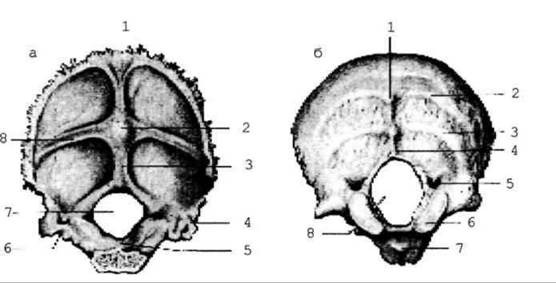
Fig. 2.8. Occipital bone: a - anterior and superior view:
On the Inferior surface of the basilar part lies the pharyngeal tubercle, and on its superior surface is the clivus. The external surface of the lateral parts features the occipital condyle, condylar fossa (or canal), jugular process, and jugular notch; the groove for the sigmoid sinus is located on their internal surface. The hypoglossal canal passes through the lateral part. The external surface of the occipital squama exhibits the external occipital protuberance and nuchal lines (highest, superior, and inferior). Its internal surface presents the internal occipital protuberance, cruciform eminence, and grooves for the superior sagittal, transverse, and sigmoid sinuses.
The Parietal bone (os parietale) is a quadrilateral curved bone plate featuring four borders, four angles, an internal surface, and an external surface (Fig. 2.9).
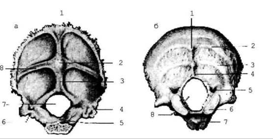
Fig. 2.9. Parietal bone
It distinguishes frontal, occipital, squamous, and sagittal borders; frontal, occipital, sphenoidal, and mastoid angles. The external surface bears the superior and inferior temporal lines, while the internal surface exhibits the grooves for the superior sagittal and sigmoid sinuses, arterial grooves, and the parietal foramen.
The Frontal bone (os frontale) comprises the squama, paired orbital parts, and an unpaired nasal part. This pneumatic bone contains the frontal sinus, inflammation of which is known as "frontal sinusitis" (Fig. 2.10).
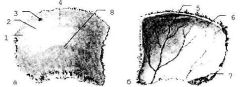
Fig. 2.10. Frontal bone: a - anterior view: 1 - frontal squama; 2 - frontal eminence; 3 - temporal line; 4 - zygomatic process; 5 - supraorbital margin; 6 - nasal part; b - posterior and inferior view: 1 - groove for the superior sagittal sinus; 2 - zygomatic process; 3 - orbital part; 4 - supraorbital notch; 5 - opening (apperture) of the frontal sinus; 6 - fossa for the lacrimal gland; 7 - frontal crest.
The nasal part features the nasal spine, ethmoidal notch, and frontal sinus. The orbital part contains the fossa for the lacrimal gland and the trochlear fossa. On the external surface of the frontal squama are the frontal eminence, superciliary arch, glabella, and supraorbital margin and foramen (or notch). The temporal surface of the squama presents the temporal line and zygomatic process. The internal surface of the squama bears the groove for the superior sagittal sinus and the foramen cecum.
The Ethmoid bone (os ethmoidale) is deeply situated within The Skull and contributes to The formation of the Nasal cavity and orbits. It consists of perpendicular, orbital, and cribriform plates. Flanking the perpendicular plate are the ethmoidal labyrinths, which contain air-filled ethmoidal Cells. The superior and middle nasal conchae hang down from the inner surface of the labyrinths into the nasal cavity (Fig. 2.11). The cribriform plate features the crista galli and ethmoidal foramina.
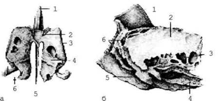
Fig. 2.11. Ethmoid bone: a - posterior view: 1 - crista galli;
2 - cribriform plate; 3 - orbital plate; 4 - superior nasal concha;
5 - perpendicular plate; 6 - ethmoidal labyrinth;
b - lateral view: 1 - crista galli; 2 - orbital plate; 3 - posterior ethmoidal cells; 4 - middle nasal concha; 5 - perpendicular plate; 6 - anterior ethmoidal cells.
The Sphenoid bone (os sphenoidale) is located between the occipital and frontal bones at the Base of the neurocranium. It consists of a body and three pairs of processes: greater wings, lesser wings, and pterygoid processes (Fig. 2.12). The superior orbital fissure, through which Blood Vessels and nerves pass, is situated between the greater and lesser wings.
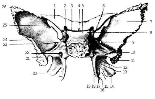
Fig. 2.12. Sphenoid bone; posterior view.
The greater wings feature cerebral, orbital, maxillary, and temporal surfaces; the latter bears the infratemporal crest. At the base of the greater wings lie three apertures: the foramen rotundum, foramen ovale, and foramen spinosum. Nerves exit the cranial cavity through the foramen rotundum and foramen ovale, whereas an artery enters the cranial cavity via the foramen spinosum.
The optic canal runs through the base of the lesser wing.
The pterygoid process features lateral and medial plates, alongside the pterygoid and scaphoid fossae, as well as the pterygoid canal and notch.
The superior surface of the body bears a depression known as the sella turcica, which consists of the hypophysial fossa and the dorsum sellae. It houses the Pituitary Gland, a vital endocrine organ. The body also contains the sphenoidal sinus (Fig. 2.13).
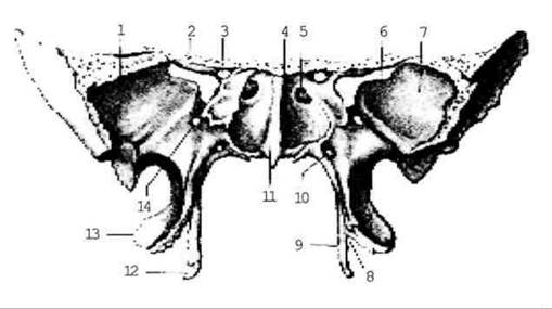
Fig. 2.13. Sphenoid bone; anterior view
The Temporal bone (os temporale) is a paired bone of complex Structure that forms part of the base and lateral wall of the cranium. It comprises three parts: 1) the squamous part, pars squamosa; 2) the tympanic part, pars tympanica; and 3) the petrous part, pars petrosa. The temporal bone contains the middle and Inner ear (Fig. 2.14).
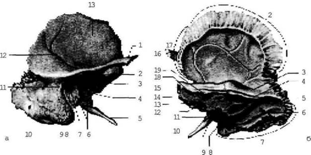
Fig. 2.14. Temporal bone; a — internal surface, b — external surface.
The squamous part, pars squamosa, contributes to the Formation of the lateral walls of the cranium. It is a vertically oriented, outwardly convex plate with a rounded margin that overlaps the corresponding edge of the parietal bone like fish scales. The zygomatic process projects from the squamous part and articulates with the zygomatic process of the Zygomatic bone to form the zygomatic arch. The squamous part presents cerebral and temporal surfaces. The latter features the groove for the middle temporal artery, the mandibular fossa, and the articular tubercle.
The tympanic part, pars tympanica, is a small, curved plate that bounds the external acoustic meatus on three sides. This meatus continues as the external auditory canal. In newborns, the tympanic part and external auditory canal are not yet fully formed, which accounts for the high incidence of tympanic cavity infections in early childhood. It is demarcated from the other parts by the tympanomastoid, tympanosquamous, petrotympanic, and petrosquamous fissures.
The petrous part (pyramid), pars petrosa, is shaped like a three-sided pyramid. It presents three surfaces—inferior (visible on the external cranial base), anterior, and posterior (both facing the cranial cavity)—along with three borders: superior, posterior, and anterior. The external relief of the pyramid is complex due to its role as a container for the Middle ear (tympanic cavity) and inner ear (osseous and membranous labyrinths), as well as the passage of nerves and vessels.
The anterior surface of the pyramid features the tegmen tympani and the trigeminal impression.
The posterior surface of the pyramid presents the internal acoustic opening, which leads into the internal acoustic meatus.
The inferior surface of the petrous part bears the jugular fossa, the petrosal fossa, the stylomastoid foramen, and the external carotid opening.
The base of the pyramid continues into the mastoid process (processus mastoideus), which contains air cells communicating with the tympanic cavity (Fig. 2.15).
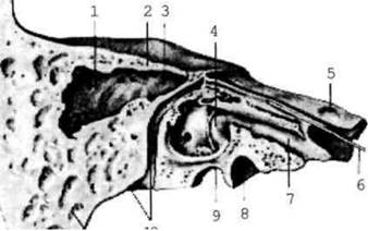
Fig. 2.15. Temporal bone; vertical section parallel to the axis of the pyramid
The pyramid of the temporal bone contains canals and canaliculi through which Blood Vessels and nerves pass. The largest is the carotid canal, transmitting the Internal Carotid Artery. It begins with its external opening on the inferior surface of the pyramid and opens internally near the apex of the pyramid.
The facial canal begins deep within the internal acoustic meatus and terminates at the stylomastoid foramen located on the inferior surface of the pyramid.
The musculotubular canal begins near the apex of the pyramid and extends toward the tympanic cavity. It is divided into two semicanals: the semicanal for the pharyngotympanic tube and the semicanal for the tensor tympani Muscle.
The caroticotympanic canaliculi originate on the wall of the carotid canal and terminate in the tympanic cavity.
The canaliculus for the chorda tympani begins above the stylomastoid foramen and opens into the tympanic cavity.
The mastoid canaliculus originates in the jugular fossa and opens into the tympanomastoid fissure.
The tympanic canaliculus begins in the petrosal fossa and ends at the hiatus for the lesser petrosal nerve.
Last update: 08/08/2026
Editorial and Educational Adaptation: This material has been compiled based on the primary/original source text. The project team performed an editorial review, corrected technical inaccuracies, structured sections, and adapted the content for an educational format.
What was processed:
- elimination of formatting defects (OCR errors, structural breaks, corrupted characters);
- editorial organization of content;
- standardization of terminology in accordance with academic sources;
- verification of factual statements against the original source text.
All mentions of the author, publication year, and origin of the primary text have been preserved in accordance with the source.