BOTANY VOLUME 1 - CELL BIOLOGY. ANATOMY. MORPHOLOGY - 2007
5. STRUCTURAL PRINCIPLES OF THALLOPHYTES
5.2. Cellular and Siphonous Thalli
The general phylogenetic trend toward increasingly large organisms manifested itself in completely different ways among thallophytes.
The seemingly simplest path of enlarging a single uninucleate Cell—i.e., The Development of macrourity—is an obvious evolutionary dead end. Plant organisms with this Structure are found in only one group of green Algae, the Dasycladaceae, which includes the well-known Acetabularia, growing to over 5 cm. Their complex vegetative bodies (see Fig. 11.92) initially contain only a single, unusually large "primary" Nucleus with a diameter of over 70 µm, from which more than 10,000 small "secondary" nuclei arise during further development.
The enlargement of a uninucleate cell is limited by the nucleocytoplasmic ratio. Especially with very large nuclei, The ratio of the nuclear surface area to its volume is unfavorable. The number of nuclear pores, whose density on the nuclear surface cannot exceed 80 per 1 µm2, is far too small relative to the volume of The Nucleus. Furthermore, the distances for Intracellular Transport of Gene products to distantly located PARTS OF THE Cytoplasm and Organelles are far too great. The second difficulty can be circumvented by The formation of multinucleate giant Cells known as coenocytes. Multinucleate, plasmodial large cells are widespread in various algal groups and in Fungi, representing the siphonaceous type of Organization (from Greek siphon, Water-Skin/tube). Thus, there are siphonaceous green algae (e.g., Caulerpa, see Fig. 11.91) and siphonaceous Xanthophyceae (Botrydium, Vaucheria, see Fig. 11.73). Among fungi, multinucleate thalli are particularly characteristic of Oomycota, Chytridiomycota, and Zygomycota.
However, this type of thallus enlargement also has its limits. On the one hand, further differentiation via the modulation of gene activity in multinucleate cells is hardly possible; on the other hand, the siphonoblast, whose single Cell wall cannot be sufficiently thick, is easily vulnerable and lacks sufficient mechanical strength. Characteristically, in the thallus of Caulerpa (see Fig. 11.91, A), which exceeds a decimeter in size, opposing parts of The Cell wall are reinforced by "struts" that extend transversely across the interior of the cell. In other algae (Cladophora, see Fig. 11.90), the siphonaceous thallus is divided into chambers by transverse walls, but such cells are still multinucleate—representing siphonocladous organization.
5.3. Multicellular Algal Thalli
5.3.1. Filamentous Thallus
The simplest form of trichal organization can be considered the filamentous thallus—the haplonema—consisting of equal-sized, single-rowed uninucleate cells following one another (Fig. 5.4, A; from Greek trichos, Hair-like, correspondingly thrix, hair, and nema, thread). A well-known example is the green alga Spirogyra (see Figs. 2.87; 11.104, A). In this Organism, all cells along the filament are equivalent, with no differentiation. In other cases, the filamentous thallus is firmly attached at one end to a specially modified cell lacking chlorophyll—a rhizoid—as, for example, in the green alga Ulothrix (see Fig. 11.89, A). In this case, the haplonema is polar, meaning There is a rhizoidal end and an apex. Polarity is often reinforced by the restriction of cell divisions to the apical cell (Fig. 5.4, B).
Class="center">Fig. 5.4. Growth and branching of a filamentous or flat algal thallus, respectively; the axes of the mitotic spindles are indicated (after D. von Denffer)
A—filamentous thallus with intercalary growth; B—growth via an apical cell; C—same with apical-polar branching; D—dichotomous division of the apical cell through divisions occurring periodically perpendicular to the direction of previous divisions; E—subapical lateral branching via unequal division of the apical cell; F—lateral branching from segments located behind the apical cell; G—formation of a flat, tissue-like thallus through the fusion of lateral branches
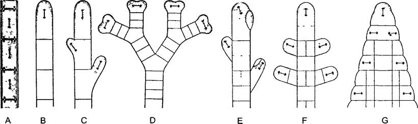
When divisions other than merely transverse ones occur in the filamentous thallus (with the spindle axis oriented longitudinally), branching takes place (Fig. 5.4, C–F). Branches can arise from the apical cell as well as from other cells. Typically, the corresponding cell divisions are unequal, leading to a hierarchical branching system: one can distinguish between the main axis and lateral branches, and upon further branching, lateral Branches of the first, second, and subsequent orders (Fig. 5.5). As a result of cell divisions proceeding in different directions, even if within a single plane, a flat thallus may eventually form. Due to its external resemblance to a leaf, it is called a phylloid (see Figs. 5.4, G; 5.8, A).
Fig. 5.5. Long SHOOT of the brown alga Halopteris filicina (after K. Goebel).
During unequal divisions, the apical cell cuts off segments that subsequently divide by transverse and longitudinal walls. Alternating with segment formation, initials that form lateral branches are separated from the apical cell by oblique, curved walls (40x)
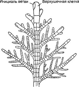
Fig. 5.6. Thallus of a red alga, central-filament type, example Chondria tenuissima (after Falkenberg): A—longitudinal section; B—cross section
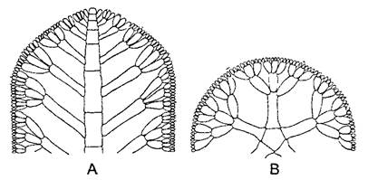
Fig. 5.7. Foliose thalli of red algae (A—F. Oltmanns, B—after Rosanoff): A—apex of a thallus branch of Fucus vesiculosus [Note: 'Furcellaria fastigiata' kept as original context]; B—single-layered thallus of Metobesia growing fan-wise in a single plane due to irregular longitudinal divisions of marginal cells (45x)

The best-known example of this type of thallus is the sea lettuce Ulva lactuca, a relative of Ulothrix (see Fig. 11.89, L), whose palm-sized, lobed thalli are frequently found in the intertidal and surf zones of European marine coasts. The phylloid of Ulva consists of two cell layers. Comparison with related species shows that these correspond to the single-layered wall of a tubular thallus, which in Ulva, however, is flattened. The necessary mechanical strength is achieved through tensile-strong, thread-like ("hyphal") cells that grow inward between the two cell layers from the rhizoidal pole of the phylloid. This constitutes a simple mechanical tissue1.
1 These correspond more closely to idioblasts rather than true tissue. — Ed. note.
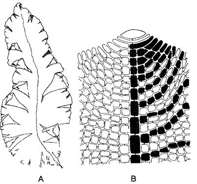
The thalli of many red algae are externally complex in their segmentation. Nevertheless, they lack true Tissues. Instead, the arrangement of cells, as an expression of ontogenetic development, is based on the filamentous thallus organization type. The vegetative bodies of most red algae are formed by numerous filaments growing in a coordinated manner, resulting in well-defined macrostructures. Individual filaments predominantly grow apically. They branch either subapically, resulting in a whorl of lateral filaments (central-filament type, Fig. 5.6), or apically (plate-like type, Fig. 5.7). Flat thalli can also arise in this manner (Fig. 5.8). In both cases, the filaments either fuse (or cement) together into a false tissue, or pseudoparenchyma, which in the mature state is hardly distinguishable from typical parenchyma, or the filaments form a dense entanglement known as plectenchyma (Greek plectos, woven). In this structure, only the cells of a single filament are interconnected by pit-connection structures.
Fig. 5.8. Leaf-like thallus of the red alga Gunnellia americana (0.5x) (A) and the apical end of a single-layered thallus (B) with a large apical cell and the central filament formed by it; the latter and some cell lines originating from it are blackened (300x) (A—after R.L. Smith, B—after J. Tilden)
5.3.2. Tissue Thalli
Among algae, the highest degree of thallus differentiation is achieved, on the one hand, by charophytes (Charophyceae, Fig. 5.9, 10, 106), and on the other, by brown algae. Marine brown algae include the largest and longest-lived aquatic plants. The thalli of certain kelps can exceed 100 m in length1. They are differentiated into holdfasts (despite being multicellular, these are still referred to as rhizoids), stem-like Organs called cauloids (from the Greek kaulos – stem), and leaf-like assimilators known as phylloids. This is accompanied by a relatively rich cellular differentiation, allowing for the distinction of epidermal (protective), cortical, and medullary tissues.
1 Such massive sizes are characteristic of brown algae from several genera, whereas true kelps are significantly smaller. — Ed. note.
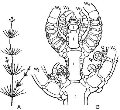
Fig. 5.9. Thallus STRUCTURE OF THE charophyte alga Chara fragilis (A – after A. W. Haupt, B – after J. Sachs)
A – segmentation into nodes with whorled branches and the internodes connecting them; a lateral shoot may form at each node (0.5x). B – longitudinal section through the thallus apex showing the apical cell. The cells cut off from it divide again unequally into a distal nodal cell and a basal internodal cell, which becomes covered with a cortex derived from the nodal cell. The internodal cells are unusually large in the mature state, exhibiting rapid cytoplasmic streaming. Branches of the whorl (W3–W5) develop from the outer nodal cells, which are likewise segmented into nodes and internodes, bearing oogonia and antheridia at their nodes (30x); 1 – internodal cells, O – oogonium, S – apical cell, Sp – spermatogonium.
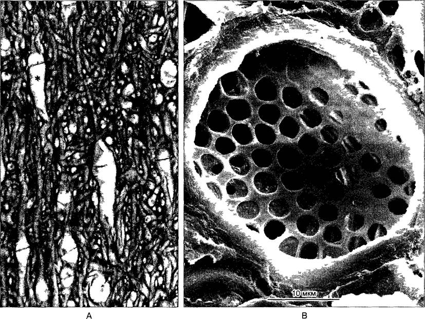
The cauloids contain so-called sieve hyphae, which structurally and functionally correspond to the sieve tube elements of angiosperms. The pore diameter in their transverse walls, which resemble sieve plates, reaches up to 6 μm (Fig. 5.10). The similarity between the sieve hyphae of brown algae and the sieve tube elements of higher plants (see Fig. 3.22) represents an analogy and serves as a striking example of convergence.
Fig. 5.10. A – entanglement of cellular filaments in the cauloid of the brown alga Laminaria, showing wide-lumen sieve hyphae (one indicated by an asterisk) with transverse sieve plates (150x). B – sieve plate in the plectenchyma of the brown alga Macrocystis integrifolia (SEM micrograph by K. Schmitz).
Last update: 07/08/2026
Editorial and Educational Adaptation: This material has been compiled based on the primary/original source text. The project team performed an editorial review, corrected technical inaccuracies, structured sections, and adapted the content for an educational format.
What was processed:
- elimination of formatting defects (OCR errors, structural breaks, corrupted characters);
- editorial organization of content;
- standardization of terminology in accordance with academic sources;
- verification of factual statements against the original source text.
All mentions of the author, publication year, and origin of the primary text have been preserved in accordance with the source.