BOTANY VOLUME 1 - CELL BIOLOGY. ANATOMY. MORPHOLOGY - 2007
5. PRINCIPLES OF ORGANIZATION IN THALLOPHYTES
5.4. Fungal Mycelium
The vegetative bodies of most Fungi (and consequently of all Lichens) are composed of segmented cellular filaments called hyphae (from Greek hyphe — web). Hyphae grow exclusively apically; only at the tip, in hyphae less than 20 µm long, is The Cell wall sufficiently soft and pliable—due to the continuous incorporation of new material—to stretch under turgor pressure. Fungal hyphae can grow very rapidly, up to 1 mm/h. Rapid growth and frequent branching within a substrate (soil, decomposing foliage, tree trunks, etc., as well as in liquids) lead to The formation of a loose network of filaments with an enormous total surface area known as the mycelium (from Greek mykes — mushroom, myzein — to suck; Fig. 5.11). Hyphae lack a cuticle, which makes the mycelium prone to drying out, but renders it exceptionally well-suited for the osmotrophic absorption of dissolved nutrients. Many higher plants exploit this by forming a Symbiosis with fungi known as mycorrhiza (see 9.2.3). However, for the exact same reasons, fungi readily accumulate toxic heavy metals (such as cadmium) and radionuclides, a factor that must be taken into account given current levels of environmental pollution.
Class="center">Fig. 5.11. Fungal mycelia (SEM micrographs: A — L. Schreiber; B — V. Kern): A — loose mycelium On the surface of a first-year fir needle (62x); B — plectenchyma of the velvet shank mushroom Flammulina velutipes (Basidiomycota) from the Physalacriaceae family

While the hyphae of lower fungi lack cross-walls and are polyenergid, meaning they represent a siphonous level of Organization, the hyphae of Ascomycetes and Basidiomycetes are divided into Cells by transverse walls called septa, thereby exhibiting a cellular Structure (see Fig. 11.43). The septa, however, feature central pores ranging from 50 to 500 nm in diameter, ensuring that the Cytoplasm remains continuous throughout the entire blastema in higher fungi as well. During the formation of short-lived fruiting bodies—commonly referred to as mushrooms—the mycelium condenses into a typical plectenchyma (see Fig. 5.11, B). In this process, specialized Tissues such as conducting or mechanical tissues are typically not differentiated, though the spore-forming hymenium often differs markedly from the rest of the plectenchyma. By contrast, the long-lived fruiting bodies of polypores contain mechanical thick-walled, elongated, and usually branched skeletal hyphae, interwoven with rigid yet short binding hyphae (see Fig. 11.58).
5.5. Organizational Forms of Liverworts and Mosses
Most mosses are terrestrial inhabitants of humid habitats. Particularly lush moss vegetation is found in tropical rain forests and foggy montane forests, in shaded places and along watercourses, as well as in fens and bogs. The presence of mosses is, in a sense, an indicator of high humidity. They require liquid Water droplets for Fertilization, as their male Gametes (spermatozoa, produced in antheridia) are flagellated and swim to the egg cells located within the archegonia. Furthermore, unicellular or multicellular rhizoids penetrate the soil by only a few millimeters and are thus unable to reach deeper water tables (mosses lack true roots). Finally, their capacity to transport water to aerial Organs is also quite limited.
Fig. 5.12. Forking branching of thalli in brown Algae (A–D) and liverworts (E, F) (A — after H. Schenk; B–D — after de Wildeman; E — after W. Kingmüeller; F — after L. Kny).
While the brown alga Dictyota dichotoma (A, 0.5x) exhibits true dichotomous branching resulting from the division of an apical cell (B–D, 250x), in liverworts (e.g., Riccia rhenana, E, 2.5x) thallus branching is initiated by the de novo formation of a second, side-by-side bilateral apical cell S2 adjacent to the existing S1 cell (F, 370x)
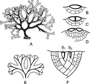
Fig. 5.13. SHOOT apex of the moss Fontinalis antipyretica (A — after H. Leitgeb; B — after O. Stocker): A — longitudinal section (120x); B — top view; the three-sided apical cell is shaded. Each segment produced by it is divided by a periclinal wall into an inner and an outer (cortical) cell. The latter forms the cortical tissue and the leaf. Lateral branches originate below a leaf through the formation of a new three-sided apical cell z. In Fontinalis, an aquatic plant submerged in flowing water, the leaves are arranged in three longitudinal rows. In most other mosses, the leaves are slightly asymmetric, resulting in a alternate spiral arrangement

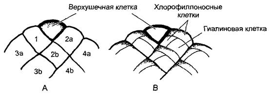
Among liverworts, the thalli reach a high level of differentiation. In many species, multi-layered tissue thalli are flat and grow via bilateral apical cells (Fig. 5.12, E, F). Their forking branching occurs not through equal division of the apical cell itself (as seen, for example, in brown algae—true dichotomy), but through the Transformation of a cell segmented from the original apical cell into a new, secondary apical cell. In so-called thallose liverworts, such as Marchantia (see Fig. 11.111), the thalli lie flat on the soil. Antheridiophores and archegoniophores rise on vertical stalks more than 1 cm high above the thallus. In many leafy liverworts, instead of a flat, lobed thallus, creeping leafy shoots are formed that are only a single cell layer thick and lack a midrib (the stem and leaves of liverworts are not homologous to the much more complex stems and leaves of cormophytes). The organization of mosses (bryopsida) is even more advanced. Their shoots grow via a three-sided apical cell1 (Fig. 5.13), and their stems possess a complex tissue structure featuring a central conducting strand (central strand, Fig. 5.14, 1), branches of which often extend into the leaves as a midrib. Leaf structure can be further elaborated through the regular alternation of equal and unequal cell divisions (Sphagnum, Figs. 5.15; 11.120); an increase in leaf surface area is also possible via the formation of longitudinal ridges on the upper surface composed of cells exceptionally rich in METABOLISM/14.html">Chloroplasts (see Fig. 11.122, A).
1 An identical cell is found at the shoot apex of leafy liverworts. — Ed. note.
Fig. 5.14. Cross-section of the stem of the moss Mnium undulatum (90x) (after E. Strasburger): a — protective tissue; I — central conducting strand; p — cortical parenchyma; rh — rhizoids
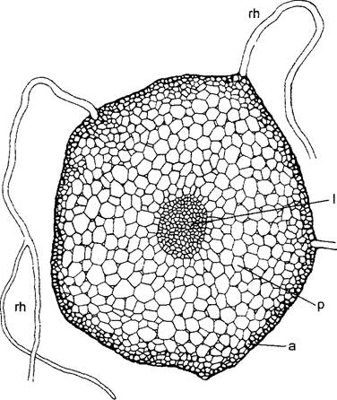
Fig. 5.15. Cell divisions in the leaf of the peat moss Sphagnum (B — after E. Bünning): A — a bilateral apical cell cuts off segments to the left and right (1–4), which subsequently divide again into identical rhombic cells (2a, 2b, 3a, 3b...); B — after apical cell divisions cease, each rhombic cell undergoes two unequal divisions to form two chlorophyllose (green) cells and one hyaline cell (150x). The hyaline cells die off after their walls are reinforced with spiral thickenings and develop a large pore opening outward; these hyaline cells serve for water storage (see Figs. 11.120, G, H)
Mosses, which are close in their organization to cormophytes, uniquely pass through various types of thallophytic organization during their development. Spore germination initiates growth at the trichal level of organization, forming a protonema — a network of uniseriate, branched cellular filaments (haplonema). Haploid gametophytic shoots producing sex cells develop on this filamentous thallus. From fertilized zygotes arise diploid sporophytes, which bear simple Stomata (located on the sporophyte, not the leaves).
Last update: 07/08/2026
Editorial and Educational Adaptation: This material has been compiled based on the primary/original source text. The project team performed an editorial review, corrected technical inaccuracies, structured sections, and adapted the content for an educational format.
What was processed:
- elimination of formatting defects (OCR errors, structural breaks, corrupted characters);
- editorial organization of content;
- standardization of terminology in accordance with academic sources;
- verification of factual statements against the original source text.
All mentions of the author, publication year, and origin of the primary text have been preserved in accordance with the source.