MEDICAL BOTANY - A.G. Serbin - 2003
CHAPTER 1. ANATOMY
PLANT TISSUES
SECRETORY TISSUES
Secretory structures ensure the synthesis, accumulation, and secretion of metabolic products such as resins, balsams, Essential Oils, saponins, rubber, mucilage, and others. Secretory structures are divided into two types: exogenous, or external (outer) secretion structures, which release secretions into the external environment, and endogenous, or internal secretion structures, which accumulate secretions or release them into adjacent Tissues.
Exogenous Secretory Structures
External secretion of substances is carried out by glandular hairs, glands, scales, nectaries, osmophores, and hydathodes.
Glandular hairs, glands, and scales (Fig. 1.17, 1–8) secrete products (essential oils, balsams, resins, etc.) first into the space between The Cell wall and the cuticle of the secretory or epithelial Cells, and subsequently release them into the atmosphere after the cuticle ruptures.
Class="center">Fig. 1.17. External secretory structures: 1–6 — glandular hairs: 1 — geranium; 2 — sage; 3 — belladonna; 4 — henbane; 5 — rosemary; 6 — foxglove; 7, 8 — essential oil glands (a — top view, b — side view): 7 — Asteraceae type; 8 — Lamiaceae type; 9 — globe thistle nectary
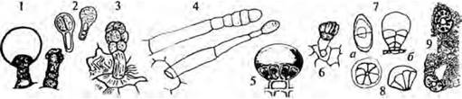
Nectaries (Fig. 1.17, 9) and osmophores are multicellular, morphologically diverse, and species-specific structures located on flowers, and more rarely on leaves. They produce a sweet liquid nectar that attracts pollinators. Sometimes petals, stamens, or other floral parts transform into nectaries. Secretion in nectaries can be performed not only by the epidermis, but also by underlying parenchyma layers.
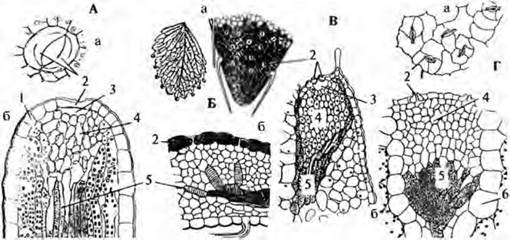
Hydathodes, or Water Stomata (Fig. 1.18), are adaptations for guttation — The excretion of weak mineral solutions, or more rarely organic solutions, in the form of droplets. Hydathodes are usually located on the Teeth of leaf margins.
Fig. 1.18. Hydathodes: A — primrose; B — cinquefoil; C — saxifrage; D — stonecrop: 1 — epidermis, 2 — guard Cells of the water stoma; 3 — water-storage cavity, 4 — epithem; 5 — tracheids; 6 — parenchymatous sheath (a — top view, b — sectional view)
Endogenous Secretory Structures
Internal secretion is carried out by idioblast cells, secretory cavities, and laticifers, which can be found in any Organs and PARTS OF THE plant.
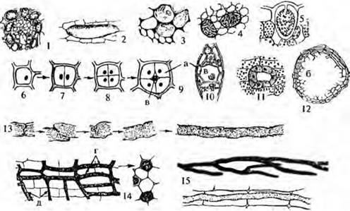
Secretory cells, or idioblasts (Fig. 1.19, 1–5), vary in shape, size, and coloration. They accumulate balsams, resins, oils, Tannins, Gums, calcium oxalate crystals, rubber, mucilages, and other substances.
Secretory cavities are cavities, canals, or ducts filled with secretions. Depending on their mode of formation, Three types of cavities are distinguished. Schizogenous cavities, ducts, and canals (Fig. 1.19, 9–11) represent intercellular spaces with clearly defined inner boundaries or tubular structures lined on the inside with secretory cells. Lysigenous cavities (Fig. 1.19, 12) are formed by the lysis of cell walls and the partial or complete breakdown of secretory cells, As a result of which the cavity boundaries are indistinct. Schizolysigenous cavities are initially formed as lysigenous ones, after which epithelial cells develop around them, or vice versa — they originate as schizogenous cavities, and subsequently the secretory cells undergo lysis.
Laticifers (Fig. 1.19, 13–15) are prosenchymatous cells or tubular articulated structures containing white or colored milky juice, known as latex. Its composition is complex, variable, and species-specific. It often contains physiologically active substances such as Alkaloids, Glycosides, resins, tannins, etc. Depending on their Structure and origin, laticifers are classified as articulated or non-articulated. Articulated laticifers form at any developmental stage of any plant organ from a vertical row of cells whose transverse walls may completely or partially persist, perforate, or break down. Lateral branches, or anastomoses, may appear, connecting neighboring latex tubes into a single system. Articulated laticifers with anastomoses are found in plants of the families Campanulaceae, Papaveraceae, and Liliaceae, while articulated laticifers without anastomoses occur in Fabaceae, Asteraceae, Alliaceae, and others. Non-articulated laticifers originate in the embryo or later, grow together with the plant, permeate all its organs, and may be branched or unbranched. Unbranched non-articulated laticifers are characteristic of Apocynaceae, Urticaceae, and Moraceae, whereas branched non-articulated laticifers are typical of Euphorbiaceae, Asclepiadaceae, and others.
Fig. 1.19. Internal Secretory Structures: 1–5 — idioblast cells: tanniniferous cell in a beech leaf, mucilage cell in a marshmallow ROOT, essential oil cell in a geranium leaf, crystal-bearing cell in a belladonna leaf, cystolith-bearing cell in a ficus leaf; 6–9 — formation of a schizogenous cavity; 10 — schizogenous resin duct in pine wood; 11 — essential oil canal in parsley root; 12 — lysigenous cavity in a mandarin pericarp; 13 — formation of an articulated laticifer without anastomoses; 14 — articulated laticifers with anastomoses (in longitudinal and transverse sections); 15 — non-articulated laticifers: branched and unbranched; a — secretory cells; b — degraded cells; c — cavity with secretion; d — latex tubes; e — anastomoses
Last update: 07/08/2026
Editorial and Educational Adaptation: This material has been compiled based on the primary/original source text. The project team performed an editorial review, corrected technical inaccuracies, structured sections, and adapted the content for an educational format.
What was processed:
- elimination of formatting defects (OCR errors, structural breaks, corrupted characters);
- editorial organization of content;
- standardization of terminology in accordance with academic sources;
- verification of factual statements against the original source text.
All mentions of the author, publication year, and origin of the primary text have been preserved in accordance with the source.