BIOLOGY Volume 1 - A Guide to General Biology - 2004
3. CHEMICAL COMPONENTS OF LIVING ORGANISMS
3.6. DNA and RNA: Nucleic Acids
3.6.3. DNA Structure
Like Proteins, Nucleic Acids possess a Primary Structure (which refers to their nucleotide sequence) as well as a three-dimensional structure. Interest in Introduction/20.html">DNA Structure surged in the early 20th century when it was first hypothesized that DNA might serve as the genetic material. We will examine the evidence supporting this role of DNA in Chapter 23.
In the early 1950s, the American chemist and Nobel laureate Linus Pauling, who had already elucidated the α-helical structure characteristic of many Fibrous proteins, turned his attention to The structure of DNA, which available evidence suggested was also a fibrous molecule. Concurrently, at King's College London, Maurice Wilkins and Rosalind Franklin were attempting to solve the same problem using X-Ray Diffraction Analysis. Their research required painstaking and time-consuming preparation of pure DNA salt samples, from which complex diffraction patterns could be obtained (Fig. 3.41). However, these patterns only revealed the general architecture of the DNA molecule rather than the high-resolution details obtainable from pure protein crystals.
Class="center">
Fig. 3.41. X-ray diffraction pattern of a DNA fiber. Such diffraction patterns first led to the Conclusion that DNA has a double-helical structure. (Courtesy of J. M. Squire.)
Meanwhile, at the Cavendish Laboratory of the University of Cambridge, James Watson and Francis Crick adopted a different approach that ultimately led to the successful solution of the problem. Drawing upon all available Physical and Chemical data, Watson and Crick began constructing three-dimensional models of DNA in the hope of eventually converging on a compelling structure consistent with all the experimental evidence. The story of their quest is vividly recounted by Watson in his book The Double Helix.
Two key factors proved decisive for Watson and Crick. First, they had regular access to Wilkins's findings, allowing them to test their models by comparing them against his X-ray diffraction images. Wilkins's X-ray data provided compelling evidence for a helical structure (Fig. 3.41) with a 0.34 nm axial periodicity. Second, Watson and Crick recognized the profound Significance of the rules governing the proportions of different bases in DNA. These regularities had been discovered and reported in 1951 by Erwin Chargaff; however, despite its immense importance, this discovery initially failed to attract the attention it deserved. Table 3.10 presents some of Chargaff's data, supplemented by the results of later research.
Table 3.10. Relative amounts of bases in DNA from various organisms
Nucleotide composition, mol % |
||||
adenine |
guanine |
thymine |
cytosine |
|
Human Sheep Chicken Turtle Salmon Sea urchin Locust Wheat Escherichia coli (bacterium) Bacteriophage φX174 (virus) |
30.9 29.3 28.8 29.7 29.7 32.8 29.3 27.3 31.3 24.7 24.6 |
19.9 21.4 20.5 22.0 20.8 17.7 20.5 22.7 18.7 26.0 24.1 |
29.4 28.3 29.2 27.9 29.1 32.1 29.3 27.1 32.9 23.6 32.7 |
19.8 21.0 21.5 21.3 20.4 17.3 20.7 22.8 17.1 25.7 18.5 |
3.13. Examine the table. What conclusion can be drawn from it regarding the proportions of different bases in DNA molecules?
Watson and Crick set out to test the hypothesis that the DNA molecule consists of two polynucleotide chains held together by base pairing between adjacent chains, with the bases stabilized by Hydrogen Bonds.
3.14. If this model is correct, can Chargaff's data be used to predict which bases pair with one another?
The hydrogen bonding between complementary bases is illustrated in Fig. 3.42. Adenine pairs with thymine, and guanine with cytosine; the AT pair is held together by two hydrogen bonds, while the GC pair is linked by three. Reflecting on how he envisioned this base-pairing arrangement, Watson later recalled: "I was thrilled to see the answer to the riddle that had been plaguing us: why the number of purine residues precisely equals the number of pyrimidine residues."1 Watson realized that this specific combination allows the bases to fit together with high precision, maintaining identical overall size and shape for both Base Pairs since each pair consists of three rings (Fig. 3.42). While hydrogen bonding between other base combinations is theoretically possible, those bonds are significantly weaker. Once these principles were established, the construction of an accurate DNA model—such as the one shown in Figs. 3.43–3.45—could finally proceed.

Fig. 3.42. Base pairing: adenine with thymine, and guanine with cytosine.
STRUCTURE OF THE DNA Molecule
Watson and Crick demonstrated that DNA is composed of two polynucleotide chains. Each chain is right-handed and coiled around a common axis, forming a double helix (Fig. 3.43). The strands are antiparallel, meaning they run in opposite directions. Each chain consists of a sugar-phosphate backbone from which the bases project inward, perpendicular to the long axis of the double helix; directly opposing bases from the two strands are held together by hydrogen bonds (Fig. 3.44). The sugar-phosphate backbones of the two strands are clearly visible in the molecular model of DNA (Fig. 3.45). The distance between the backbones remains constant, corresponding to the width of one base pair—specifically, one purine and one pyrimidine. Two Purines would occupy too much space, whereas two Pyrimidines would leave too large a gap to bridge the distance between the two chains. Along the molecular axis, adjacent base pairs are spaced 0.34 nm apart, which accounts for the periodicity observed in X-ray diffraction patterns. A complete turn of the helix spans 3.4 nm and comprises 10 base pairs. There are no sequence restrictions along a single polynucleotide chain; however, due to the base-pairing rules, The sequence of one strand entirely dictates the sequence of the other. Consequently, the two strands of the double helix are said to be complementary to each other.

Fig. 3.43. Schematic representation of DNA structure. One full turn of the helix contains 10 base pairs, with a distance of 0.34 nm between adjacent base pairs.

Fig. 3.44. DNA (schematic representation with unwound strands).
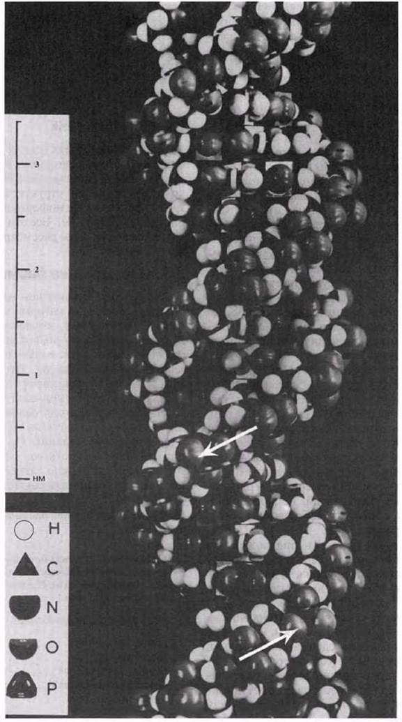
Fig. 3.45. Three-dimensional model of DNA. Arrows indicate the direction of the antiparallel sugar-phosphate backbones of the two polynucleotide chains.
Watson and Crick published their report on the DNA model in the journal Nature in 1953, and in 1962, together with Maurice Wilkins, they were awarded the Nobel Prize for this work. That same year, Kendrew and Perutz received the Nobel Prize for their work on determining the three-dimensional structure of proteins, also carried out using X-ray crystallography. Rosalind Franklin, who died of Cancer prior to the award of these prizes, was not included among the laureates because the Nobel Prize is not awarded posthumously.
To establish that the proposed structure served as the genetic material, it was necessary to demonstrate its ability to: 1) carry encoded information, and 2) reproduce (replicate) accurately. Watson and Crick were well aware that their model satisfied these requirements. At the end of their first paper, they cautiously noted: "It has not escaped our notice that the specific pairing we have postulated immediately suggests a possible copying mechanism for the genetic material"2. In a second paper published later that same year in 1953, they discussed the genetic implications of their model (which will be covered in Chapter 23). This discovery, which vividly demonstrated how structure can be linked to function at THE MOLECULAR LEVEL, gave a powerful impetus to The Development of molecular biology.
1 From The Double Helix by James D. Watson, Weidenfeld & Nicolson, 1968.
2 Watson J. D., Crick F. H. C. (1953) Nature, 171, 737.
Last update: 06/08/2026
Editorial and Educational Adaptation: This material has been compiled based on the primary/original source text. The project team performed an editorial review, corrected technical inaccuracies, structured sections, and adapted the content for an educational format.
What was processed:
- elimination of formatting defects (OCR errors, structural breaks, corrupted characters);
- editorial organization of content;
- standardization of terminology in accordance with academic sources;
- verification of factual statements against the original source text.
All mentions of the author, publication year, and origin of the primary text have been preserved in accordance with the source.