Human Biochemistry, Volume 2 - Murray R. 1993
Biochemistry of Intracellular and Intercellular Communication
Adrenal Cortex Hormones
Regulation of Adrenal Steroid Hormone Synthesis
Glucocorticoid Hormones
Cortisol secretion is regulated by ACTH, the release of which, in turn, is controlled by corticotropin-releasing hormone (CRH; corticoliberin). These hormones are linked in a classic negative feedback loop (Fig. 48.5). An elevation in free cortisol levels suppresses CRH secretion. A drop in free cortisol below normal activates the system by stimulating hypothalamic CRH release. This 41-amino-acid peptide enhances the synthesis and release of ACTH (from its precursor molecule pro-opiomelanocortin [POMC], see ch. 45). In the adrenal cortex, ACTH accelerates the side-chain Cleavage of Cholesterol—the rate-limiting step in steroidogenesis overall. These processes represent one half of the negative feedback loop. As free cortisol levels in the Blood normalize, hypothalamic CRH secretion declines, leading to reduced pituitary ACTH output and, consequently, lower adrenal cortisol production; this completes the second half of the feedback loop. This intricate mechanism ensures rapid regulation of circulating cortisol levels.
Class="center">
Fig. 48.5. Feedback regulation of cortisol Biosynthesis. Solid lines indicate stimulation; dashed lines indicate inhibition.
ACTH release (and cortisol secretion) is modulated by neural impulses originating in various parts of The Nervous system. An endogenous rhythm governs the secretion of CRH and, consequently, ACTH. This circadian rhythm is normally tuned to trigger a rise in blood cortisol shortly after Sleep onset. During sleep, cortisol levels continue to climb, peaking shortly after waking, and then gradually decline to minimal values by late evening and early night hours. This overall dynamic stems from successive episodes of pulsatile cortisol release preceded by pulsatile ACTH secretion (see ch. 45). Together, these events form a complex cycle dependent on photoperiod, feeding-fasting cycles, and sleep-wake patterns. The loss of daily steroid periodicity is typically associated with Pathologies of the pituitary-adrenal axis, certain depressive states, and transmeridian travel across multiple time zones.
Cortisol secretion is also influenced by physical and emotional stress, anxiety, fear, apprehension, and pain. These reactions can override the effects of the negative feedback system and the circadian rhythm.
Mineralocorticoid hormones
The production of aldosterone by the zona glomerulosa Cells is regulated quite differently: the primary regulators are the Renin-Angiotensin System and potassium, though sodium, ACTH, and neural mechanisms also participate in this process.
A. The renin-angiotensin system. This system plays a key role in Blood Pressure Regulation and electrolyte balance. The principal hormone here is angiotensin II, an octapeptide derived from angiotensinogen (Fig. 48.6). Angiotensinogen is an α2-globulin synthesized by the Liver; it serves as the substrate for renin, an enzyme produced by the juxtaglomerular Cells of the renal afferent arterioles. The localization of these cells makes them particularly sensitive to blood pressure fluctuations; many physiological regulators of renin release (Table 48.3) act via renal baroreceptors. Juxtaglomerular cells are also responsive to changes in Na+ and K+ concentrations in the fluid flowing through the renal tubules; consequently, any combination of factors causing fluid volume depletion (dehydration, hypotension, blood or fluid loss) or a drop in NaCl concentration stimulates renin release. Furthermore, renin release is influenced by the Central Nervous System and changes in body posture. Appropriate signals are transmitted via sympathetic nerves to the juxtaglomerular cells, acting through a mechanism independent of baroreceptors and salt effects, but mediated by ß-adrenergic receptors.
Table 48.3. Factors influencing renin release
|
Stimulating |
Inhibiting |
|
Decreased blood pressure |
Increased blood pressure |
|
Change in posture: horizontal to vertical |
Change in posture: vertical to horizontal |
|
Body salt depletion |
Salt loading |
|
ß-Adrenergic agents |
ß-Adrenergic antagonists |
|
Prostaglandin inhibitors |
|
|
Potassium |
|
|
Vasopressin |
|
|
Angiotensin II |
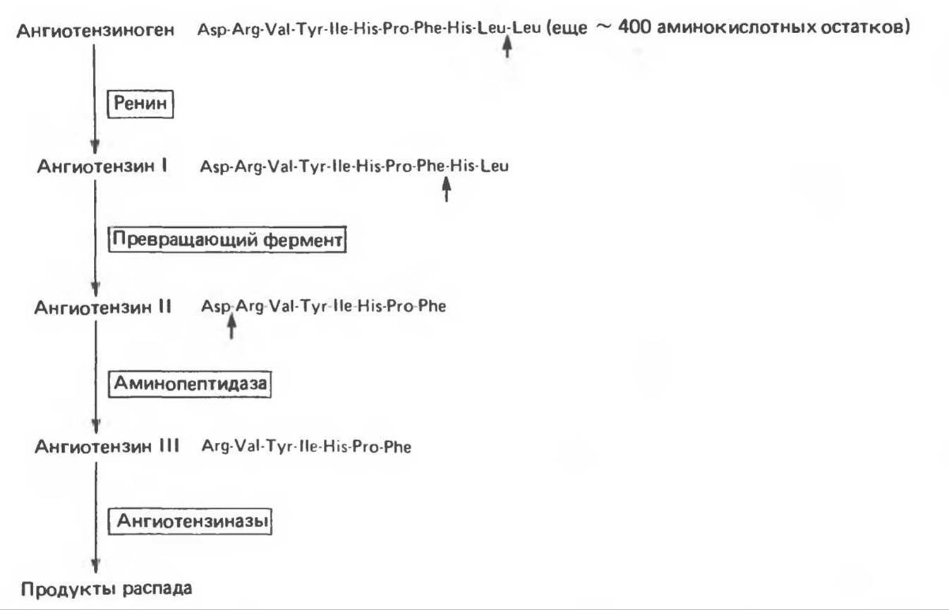
Fig. 48.6. Formation and METABOLISM of angiotensins. Cleavage sites are indicated by small arrows.
Renin acts on its substrate, angiotensinogen, converting it into the decapeptide angiotensin I. Glucocorticoids and estrogens stimulate the hepatic synthesis of angiotensinogen. The Hypertension induced by these hormones may be partly due to elevated plasma levels of angiotensinogen. Because the plasma concentration of this protein is close to the Km of its reaction with renin, minor fluctuations in this concentration can profoundly impact angiotensin II formation.
Angiotensin-converting enzyme, a glycoprotein identified in the Lungs, endothelial cells, and Blood Plasma, cleaves two C-terminal amino acid residues from angiotensin I, converting it into angiotensin II. This reaction does not appear to be rate-limiting. Various nonapeptides—analogs of angiotensin I—can inhibit the converting enzyme and are therefore used to treat renin-dependent hypertension. The converting enzyme also degrades bradykinin, a potent vasodilator. Thus, this enzyme elevates blood pressure through two distinct pathways.
Angiotensin II raises blood pressure by causing arteriolar constriction and is the most potent of all known vasoactive agents. In addition, it suppresses renin release from juxtaglomerular cells and exerts a strong stimulatory effect on aldosterone production. Although this adrenal effect is direct, angiotensin II does not influence cortisol synthesis. In some animal species, angiotensin II is converted into the heptapeptide angiotensin III (Fig. 48.6) via the removal of the Asp1 residue. The stimulatory effect on aldosterone production is roughly equivalent for both angiotensins. In humans, plasma angiotensin II levels are four times higher than those of angiotensin III, making the octapeptide the primary effector. Angiotensins II and III are rapidly inactivated by angiotensinases.
Angiotensin II binds to specific receptors on glomerulosa cells. The density of these receptors is subject to upregulation by potassium ions and the hormone itself, as well as downregulation by low potassium concentrations; thus, this ion plays a central role in the adrenal action of angiotensin II. This hormone-receptor interaction does not activate adenylate cyclase, suggesting that cAMP is not involved in the MECHANISM OF ACTION of angiotensin II.
The action of this hormone—which stimulates The conversion of cholesterol to pregnenolone and of corticosterone to 18-hydroxycorticosterone and aldosterone—may be mediated by changes in intracellular calcium concentration and phospholipid metabolites via a mechanism similar to that described in ch. 44. Prostaglandin biosynthesis may also play a role, given that prostaglandins E1 and E2 stimulate aldosterone release, whereas F1a and F2a inhibit it, which is generally typical of prostaglandin-mediated responses. Indomethacin, an inhibitor of prostaglandin biosynthesis, suppresses both basal and angiotensin II-stimulated aldosterone release.
B. Potassium. Aldosterone secretion is sensitive to changes in plasma potassium levels: an increase of just 0.1 mEq/L stimulates secretion, whereas a drop of the same magnitude inhibits hormone synthesis and release. The Effect of K+ is independent of plasma Na+ and angiotensin II levels. Prolonged hyperkalemia leads to Hypertrophy of the zona glomerulosa and enhances the sensitivity of its cells to potassium ions. K+ acts on the same enzymatic steps as angiotensin II, though its exact mechanism remains unknown. Like angiotensin II, K+ does not affect cortisol biosynthesis.
C. ACTH. In humans, a rapid, short-term drop in ACTH levels (e.g., following hypophysectomy or suppression by glucocorticoids) has little impact on aldosterone production, but chronic ACTH deficiency can blunt the response of aldosterone levels to other regulators (angiotensin II, Na+, K+). In other species (such as rats), ACTH plays a more critical role in aldosterone output: studies on isolated zona glomerulosa cells have shown that it stimulates cAMP synthesis and the initial steps of steroidogenesis.
D. Sodium. Sodium deficiency enhances aldosterone production, while a sodium load suppresses it; however, these effects are largely mediated by the renin-angiotensin system. A direct effect of Na+ on aldosterone synthesis is also possible, but this effect is weak, transient, and requires high Na+ concentrations.
Last update: 06/08/2026
Editorial and Educational Adaptation: This material has been compiled based on the primary/original source text. The project team performed an editorial review, corrected technical inaccuracies, structured sections, and adapted the content for an educational format.
What was processed:
- elimination of formatting defects (OCR errors, structural breaks, corrupted characters);
- editorial organization of content;
- standardization of terminology in accordance with academic sources;
- verification of factual statements against the original source text.
All mentions of the author, publication year, and origin of the primary text have been preserved in accordance with the source.