Harper's Illustrated Biochemistry, Volume 1 - Murray R. 1993
Structure and Functions of Proteins and Enzymes
Proteins: Structure and Properties
Ordered Conformations of Polypeptides
a-Helix
A fundamental milestone in our understanding of the principles of protein Organization was the discovery that polypeptide chains can adopt highly ordered Conformations stabilized by Hydrogen Bonds between peptide groups. Although the existence of such highly ordered structures was subsequently confirmed repeatedly by high-resolution X-ray crystallography of protein crystals, the concept was initially purely theoretical.
According to X-Ray Diffraction data obtained in the early 1930s, Hair and wool a-Keratins exhibit a meridional repeat of 0.5–0.55 nm. However, such a spacing could not be accounted for by a fully extended polypeptide chain (Fig. 5.2). This apparent contradiction was resolved by Pauling and Corey, who proposed that The polypeptide chains of a-keratin are folded into an a-helix (Fig. 5.3). In this Structure, the R-groups at the a-carbon atoms point outward from the helix axis (Fig. 5.4). There are 3.6 amino acid residues per turn of the helix, and the pitch of the helix is 0.54 nm, which closely matches the 0.5–0.55 nm periodicity observed in diffraction patterns. The axial rise per residue is 0.15 nm, which is also in agreement with the X-ray data. The Main Features of the a-helix are summarized below (Fig. 5.5).
Class="center">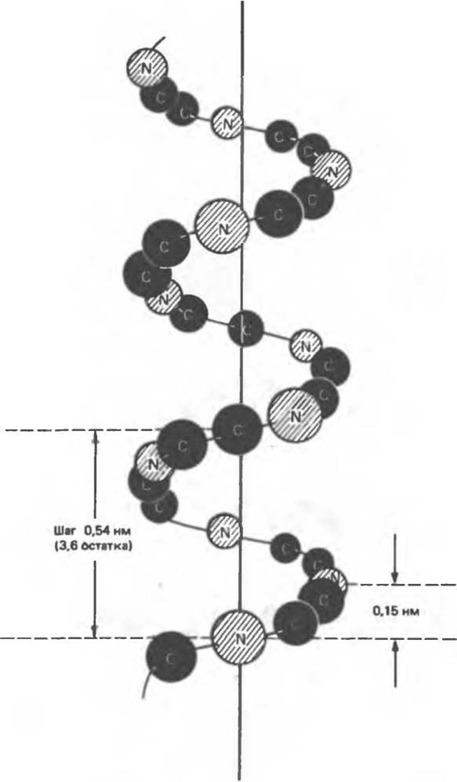
Fig. 5.3. Arrangement of the atoms of the peptide chain backbone relative to the a-helix axis.

Fig. 5.4. Top view of the a-helix. R represents side chains projecting from the helix. The Van der Waals radii of the atoms are considerably larger than shown here, leaving almost no free space inside the helix. (Reproduced with minor modifications from Stryer L., Biochemistry, 2nd ed., Freeman, 1981.)
1. The a-helix is stabilized by hydrogen bonds between the hydrogen atom attached to the nitrogen atom of a peptide group and the carbonyl oxygen of the residue situated four positions further along the chain.
2. All peptide groups participate in Hydrogen bond formation, which ensures maximum Stability of the a-helix.
3. All nitrogen and oxygen atoms of the peptide groups are involved in hydrogen bonding, which significantly decreases the hydrophilicity of the a-helical regions (and increases their Hydrophobicity).
4. The a-helix forms spontaneously and represents the most stable conformation of the polypeptide chain corresponding to a Free energy minimum.
5. In a chain composed of L-Amino Acids, the right-handed helix, which is commonly found in Proteins, is much more stable than the left-handed one.

Fig. 5.5. STRUCTURE OF THE a-helix. The a-helical conformation is largely determined by The Nature of the R-groups and is stabilized by hydrogen bonds between H and O atoms (shown as dashed lines). (Reproduced with permission from Haggis G. H. et al., Introduction to Molecular Biology, Wiley, 1964.)
Certain amino acids hinder the coiling of the chain into an a-helix, disrupting its continuity at their position. These include Proline (in which the nitrogen atom is part of a rigid ring structure, making rotation around the N — Са bond impossible), as well as amino acids with charged or bulky R-groups that electrostatically or sterically interfere with a-helix formation (Table 5.3).
ß-Pleated Sheet
Pauling and Corey proposed another ordered structure—the ß-pleated sheet (the designation ß indicated that it was the second structure they proposed after the a-helix). Whereas the polypeptide chain in the a-helix is in a compact state, the chains in the ß-pleated sheet are almost fully extended (Fig. 5.6). When adjacent polypeptide chains in a ß-pleated sheet run in opposite directions (the direction from the N-terminus to the C-terminus being taken as positive), the structure is called an antiparallel ß-pleated sheet (shown in Fig. 5.6). When adjacent chains run in the same direction, the ß-sheet structure is termed parallel (not shown in the figure).
Table 5.3. Effect of various amino acids on a-helix formation
|
formers |
disruptors |
breakers |
|
Ala |
Arg |
Pro |
|
Asn |
Asp |
Hyp |
|
Cys |
Glu |
|
|
Gln |
Gly |
|
|
His |
Lys |
|
|
Leu |
Ile |
|
|
Met |
Ser |
|
|
Phe |
Thr |
|
|
Irp |
||
|
Туr |
||
|
Val |
Regions of ß-pleated structure are found in many proteins, with both parallel and antiparallel forms occurring. From two to five adjacent polypeptide chains can participate in The formation of such structures. Fig. 5.7 shows a segment of a Ribonuclease molecule in which the ß-pleated structure is formed by three stretches of the polypeptide chain. Many proteins contain both a-helices and ß-pleated structures simultaneously (Fig. 5.7).
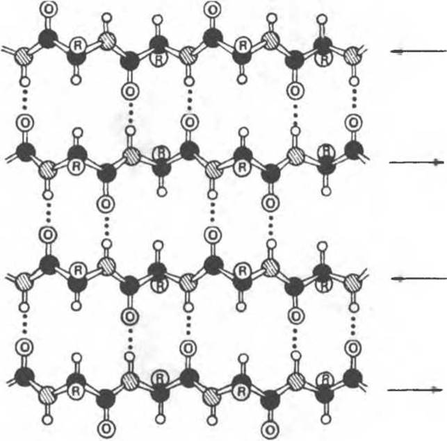
Fig. 5.6. Antiparallel ß-pleated sheet. The directions of adjacent chains are opposite. The structure is stabilized by hydrogen bonds between the NH and CO groups of adjacent chains. Side groups (R) are located above or below the plane of the sheet. Black circles represent carbon atoms, grey circles represent nitrogen atoms, and light circles represent hydrogen atoms. (Adapted from Stryer L., Biochemistry, 2nd ed., Freeman, 1981.)

Fig. 5.7. Schematic representation of chain folding in the bovine pancreatic ribonuclease molecule—a single sequence of 124 residues. The structure is stabilized by four cross-linking Disulfide Bonds. The a-helical region is outlined by an oval dashed contour, and the pleated sheet region is shaded. Other PARTS OF THE structure have an irregular conformation. The Location OF THE Active Site (Chapter 8) is indicated by the РO3-4 ion. (From Kartha G., Bello J., Harker D.: Tertiary Structure of ribonuclease. Nature 1967:213:862.) This protein has been successfully synthesized by purely chemical means.
In the a-helix, stabilizing Hydrogen bonds are formed between peptide groups spaced four residues apart along the chain, whereas the ß-pleated structure is formed through hydrogen bonds between peptide groups that are much further apart in the sequence. This fact is also illustrated in Fig. 5.7.
Disordered conformation (random coil)
Those Regions of the protein molecule that do not belong to helical or pleated structures are usually called disordered. As shown in Fig. 5.7, a significant part of the protein molecule can exist in such a conformation. The term «disordered» is not entirely appropriate, as it creates the impression of lower biological significance for these regions compared to highly ordered periodic ones. At the same time, from the perspective of biological function, disordered, irregular regions are just as important as α-helices and pleated β-sheets.
Last update: 06/08/2026
Editorial and Educational Adaptation: This material has been compiled based on the primary/original source text. The project team performed an editorial review, corrected technical inaccuracies, structured sections, and adapted the content for an educational format.
What was processed:
- elimination of formatting defects (OCR errors, structural breaks, corrupted characters);
- editorial organization of content;
- standardization of terminology in accordance with academic sources;
- verification of factual statements against the original source text.
All mentions of the author, publication year, and origin of the primary text have been preserved in accordance with the source.