Human Biochemistry, Volume 1 - Murray R. 1993
Bioenergetics and Carbohydrate and Lipid Metabolism
Oxidative Phosphorylation and Mitochondrial Transport Systems
Organization of the Respiratory Chain in Mitochondria
The Main Components of the Respiratory Chain (Fig. 13.2) are listed in order of increasing redox potential in Table 12.1. Hydrogen atoms or electrons move along the chain from more electronegative components to more electropositive oxygen; The change in redox potential during the transition from the NAD+/NADH system to the O2/H2O system is 1.1 V.
The main respiratory chain in Cell/35.html">Mitochondria begins with NAD-dependent dehydrogenases, passes through Flavoproteins and Cytochromes, and terminates with molecular oxygen. Not all substrates are linked to the respiratory chain via NAD-dependent dehydrogenases; some of them, having a relatively high redox potential (e.g., the fumarate/succinate system, see Table 12.1), are linked to flavoprotein dehydrogenases, which in turn are connected to the respiratory chain cytochromes (Fig. 13.3).
It has recently been established that the respiratory chain contains yet another carrier that links flavoproteins to cytochrome b, which has the lowest redox potential among cytochromes. This carrier, called ubiquinone or coenzyme Q (Fig. 13.4), exists in mitochondria in the oxidized quinone form under aerobic conditions, and in the reduced quinol form under anaerobic conditions. Coenzyme Q is a component of mitochondrial Lipids; Phospholipids, which form part of the mitochondrial membrane, predominate among other lipids. The Structure of coenzyme Q is similar to that of Vitamins K and E. Plastoquinone, found in METABOLISM/14.html">Chloroplasts, has a closely related structure. All these substances feature a polyisoprenoid side chain in their structure. The content of coenzyme Q significantly exceeds that of other respiratory chain components (in terms of stoichiometry); this suggests that coenzyme Q is a mobile component of the respiratory chain that receives reducing equivalents from fixed flavoprotein complexes and transfers them to cytochromes.
Class="center">
Fig. 13.1. Main sources of reducing equivalents and their connection to the mitochondrial respiratory chain. The primary extramitochondrial source is NADH, which is generated during Glycolysis.
An additional component found in functionally active preparations of the respiratory chain is the iron-sulfur protein, FeS (non-heme iron). It is associated with flavoproteins (metalloflavoproteins) and with cytochrome b. Iron and sulfur are believed to participate in the redox process proceeding via a single-electron mechanism (Fig. 13.5).
Modern concepts regarding The sequence of the main Components of the respiratory chain are reflected in Fig. 13.3. At the electronegative end of the chain, dehydrogenases catalyze The transfer of electrons from substrates to NAD located within the respiratory chain. This occurs via two pathways. In cases where the substrates are α-keto acids, Pyruvate, and ketoglutarate, complex dehydrogenase systems containing lipoate and FAD participate in transferring electrons to NAD. The transfer of electrons by Other dehydrogenases utilizing L(+)-3-hydroxyacyl-CoA, D(—)-3-hydroxybutyrate, Proline, glutamate, malate, and isocitrate as substrates proceeds directly to the respiratory chain NAD.
Reduced NADH in the respiratory chain is, in turn, oxidized by the metalloflavoprotein NADH dehydrogenase. This enzyme contains FeS and FMN and is firmly bound to the respiratory chain. Coenzyme Q serves as a collector of reducing equivalents supplied by A number of substrates via flavoprotein dehydrogenases into the respiratory chain. These substrates include succinate, Choline, glycerol-3-phosphate, Sarcosine, dimethylglycine, and acyl-CoA (Fig. 13.3). The flavin component of these dehydrogenases appears to be FAD. The flow of electrons from coenzyme Q then proceeds through a series of cytochromes to molecular oxygen (Fig. 13.3). Cytochromes are arranged in order of increasing redox potential. The terminal cytochrome aa3 (cytochrome c oxidase) carries out The final stage of the process—the transfer of reducing equivalents to molecular oxygen. As already mentioned, this enzyme system contains copper, an essential component of true oxidases. Cytochrome oxidase has a very high affinity for oxygen, allowing the respiratory chain to function at maximum rate until O2 is practically depleted in the tissue. This reaction, catalyzed by cytochrome oxidase, is irreversible; it determines the direction of movement of reducing equivalents in the respiratory chain, which is coupled with ATP formation.
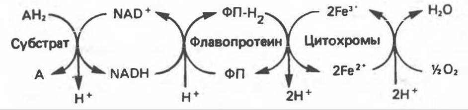
Fig. 13.2. Transport of reducing equivalents along the respiratory chain.

Fig. 13.3. Components of the mitochondrial respiratory chain. FeS is located in the chain on the "O2 side" of FP or Cyt b. Cyt — cytochrome; ETF — electron-transferring flavoprotein; FeS — iron-sulfur protein; FP — flavoprotein; Q — ubiquinone.
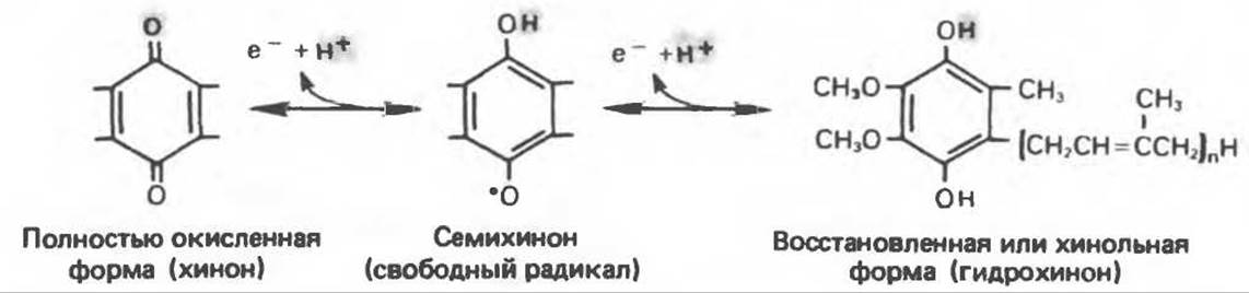
Fig. 13.4. Structure of ubiquinone (Q); n is the number of isoprenoid units, varying from 6 to 10, i.e., Q
A number of hypotheses have been put forward regarding the Structural Organization of the respiratory chain. Crucially, the molar ratios among the components are nearly constant. The functioning components of the respiratory chain are embedded in The inner mitochondrial membrane as four protein-lipid complexes of the respiratory chain. On this basis, a definite spatial orientation of these complexes within the membrane has been deduced. Cytochrome c is the only soluble cytochrome and, along with coenzyme Q, serves as a relatively mobile component of the respiratory chain, providing communication between spatially fixed complexes (Fig. 13.6).

Fig. 13.5. Iron-sulfur center (Fe4S4) of the iron-sulfur protein. S — acid-labile sulfur; Pr — apoprotein; Cys — Cysteine residue. Some iron-sulfur Proteins contain 2 iron atoms and 2 sulfur atoms (Fe2S2).

Fig. 13.6. Proposed sites of inhibition (⊖) of the respiratory chain by specific drugs, chemical Reagents, and Antibiotics. Sites where coupling with phosphorylation presumably occurs are indicated. BAL — dimercaprol; TTFA — iron-chelating reagent. Complex I — NADH:ubiquinone oxidoreductase; Complex II — succinate:ubiquinone oxidoreductase; Complex III — ubiquinol:ferricytochrome c oxidoreductase; Complex IV — ferrocytochrome c:oxygen oxidoreductase. Other Abbreviations are the same as in Fig. 13.3.
Last update: 06/08/2026
Editorial and Educational Adaptation: This material has been compiled based on the primary/original source text. The project team performed an editorial review, corrected technical inaccuracies, structured sections, and adapted the content for an educational format.
What was processed:
- elimination of formatting defects (OCR errors, structural breaks, corrupted characters);
- editorial organization of content;
- standardization of terminology in accordance with academic sources;
- verification of factual statements against the original source text.
All mentions of the author, publication year, and origin of the primary text have been preserved in accordance with the source.