Principles of Biochemistry, Volume 1 - A. Lehninger 1985
Biomolecules
Carbohydrates: Structure and Biological Functions
Some polysaccharides represent a form of storage for "cellular fuel"
The most important storage polysaccharide in plant Cells is starch, and in animal cells, it is Glycogen. Both starch and glycogen are stored inside cells as large clusters, or granules (Fig. 11-14). Starch and glycogen molecules feature many exposed hydroxyl groups and are therefore highly hydrated. When starch and glycogen are extracted from the granules with hot Water, they form turbid colloidal solutions or Suspensions.
Tubers (such as potatoes) and seeds (especially corn) are particularly rich in starch, yet The ability to synthesize starch is found in almost all plant cells (Fig. 11-14). Starch is a mixture of two glucose polymers: a-amylose and amylopectin. The former consists of long, unbranched chains of D-glucose residues linked to one another by a(1→4) bonds. The Molecular Weight of such chains ranges from several thousand to 500,000. Amylopectin also has a high molecular weight, but unlike a-amylose, its chains are highly branched (Fig. 11-15). In the unbranched regions of amylopectin, glucose residues are linked by a(1→4) bonds, whereas at the branch points, they are linked by (1→6) bonds. When potatoes are boiled, amylose is extracted by hot water, causing the water to opalesce and acquire a milky hue. In boiled potatoes, the bulk of the starch consists of the remaining amylopectin.
Class="center">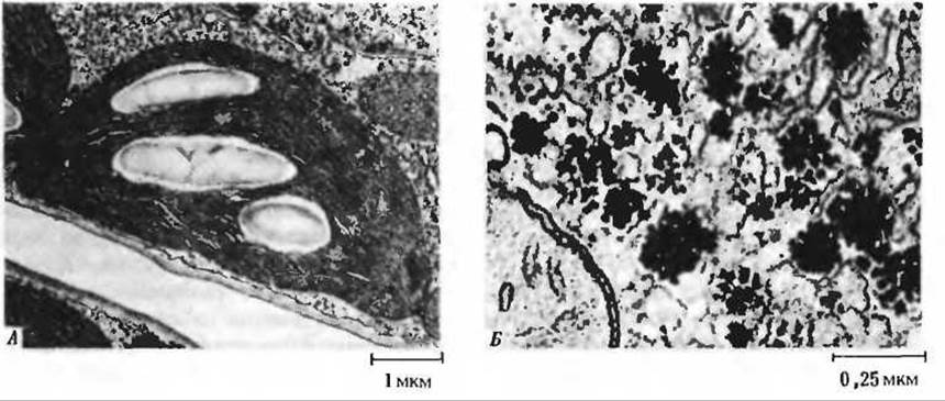
Fig. 11-14. Starch and glycogen are stored as granules in PLANT AND ANIMAL cells, respectively. A. Large starch granules in an individual chloroplast. In the leaf cells of most plants, starch is formed from D-glucose synthesized during Photosynthesis. B. Electron micrograph of glycogen granules in a hamster Liver Cell. These granules are much smaller than the starch granules shown in the adjacent image.
Glycogen is the principal storage polysaccharide in animal cells, meaning its role is analogous to that of starch in plant cells. Like amylopectin, glycogen is a branched polysaccharide consisting of D-glucose residues linked by a(1→4) bonds, but compared to amylopectin, it is significantly more branched and compact. Branch points are formed by a(1→6) bonds. Glycogen is most abundant in the liver, where it accounts for up to 7% of the organ's total weight; glycogen is also present in Skeletal Muscle. In liver cells, glycogen occurs as large granules, which in turn consist of smaller granules formed by individual, highly branched glycogen molecules with an average molecular weight of several million (Fig. 11-14). The Enzymes responsible for Glycogen Synthesis and degradation are tightly bound to these same granules.
In the gastrointestinal tract, glycogen and starch are broken down by amylases. Saliva and pancreatic juice contain a-amylases, which hydrolyze a(1→4) bonds in the outer branches of glycogen and amylopectin, releasing D-glucose, small amounts of maltose, and leaving an amylase-resistant "core" known as limit dextrin (Fig. 11-15). Dextrins are sticky substances that form the basis for preparing various adhesives. a-Amylase is unable to attack a(1→6) bonds at branch points and therefore does not hydrolyze limit dextrin; this requires a specialized enzyme, a(1→6)-glucosidase. Once a(1→6) bonds are hydrolyzed by this enzyme, another set of a(1→4) bonds becomes accessible to a-amylase. Following their Cleavage, the next set of branch points is exposed and subjected to a fresh attack by a(1→6)-glucosidase. Thus, through the concerted action of a-amylase and a(1→6)-glucosidase, glycogen and amylopectin are completely broken down to yield glucose and small amounts of maltose. In animal cells, however, glycogen is degraded by a different enzyme—namely, Glycogen phosphorylase—which cleaves glycogen to yield glucose-1-phosphate rather than free glucose (Sections 15.8 and 20.14).

Fig. 11-15. The starch Polysaccharides amylose and amylopectin. A. Amylose is a linear polymer of D-glucose residues joined by a(1→4) linkages. B. Amylopectin. Each circle corresponds to a glucose residue. Red circles denote glucose residues in the outer chains that are cleaved by the action of a-amylase. Black circles illustrate The Structure of limit dextrin formed after all outer glucose residues have been cleaved off by a-amylase. The a(1→6) bonds at chain branch points (indicated by small arrows) are hydrolyzed by a(1→6)-glucosidase, after which a new set of a(1→4) bonds becomes accessible to the amylase. Glycogen has a similar structure, but its molecule is more compact and highly branched. C. Structure of a chain branch point.
The enzyme ß-amylase found in malt differs from a-amylase in that it hydrolyzes a(1→4) bonds not consecutively, but every other bond, yielding primarily maltose and only small amounts of glucose. It should be noted that the designations a and ß in the names of amylases have nothing to do with the designations a and ß for glycosidic bonds, but are simply used to distinguish between Two Types of amylases.
Last update: 06/08/2026
Editorial and Educational Adaptation: This material has been compiled based on the primary/original source text. The project team performed an editorial review, corrected technical inaccuracies, structured sections, and adapted the content for an educational format.
What was processed:
- elimination of formatting defects (OCR errors, structural breaks, corrupted characters);
- editorial organization of content;
- standardization of terminology in accordance with academic sources;
- verification of factual statements against the original source text.
All mentions of the author, publication year, and origin of the primary text have been preserved in accordance with the source.