INVERTEBRATE ZOOLOGY IN THREE VOLUMES — BOOK 3 — H.Y. Shcherbak — 1997
PHYLUM POGONOPHORA
CLASS AFRENULATA (OR VESTIMENTIFERA)
Afrenulates are relatively large pogonophorans, reaching lengths of up to 1.5 m with a diameter of 1 cm, inhabiting the floors of seas and oceans near hydrothermal vents and cold seeps rich in hydrogen sulfide or methane (Fig. 157). The first species, Lamellibrachia barhami, was described in 1969 by the American scientist Webb, who established the new Class Afrenulata for it, while assigning all other pogonophorans known at the time to the class Frenulata. To date, 15 species of vestimentiferans are known.

Fig. 157. A colony of Riftia pachyptila specimens
The body of afrenulates, like that of other pogonophorans, consists of four regions (Fig. 158). The foremost region bears a pair of muscular outgrowths directed forward and connected by cuticular membranes to form the so-called obturaculum (from the Latin *abturo* — to plug). At the anterior end of the obturaculum lies a specialized cuticular lid that closes the tube entrance when the animal retreats inside. The obturaculum develops from larval tentacles; it contains a coelomic canal with Blood Vessels supported by a mesentery and innervated by the same nerve as the tentacles. Obturacula differ from tentacles by a greater development of Connective Tissue, the absence of pinnules, and a lack of epidermal cilia. Flanking the obturaculum are numerous (up to 300 in Riftia pachyptila) tentacular or, as they are often called, branchial lamellae, which consist of hundreds of tentacles (branchial filaments) tightly bound together by cuticle. Only their distal tips remain free; these bear pinnules, which in afrenulates are blood-filled outgrowths of the tentacular wall supplied with
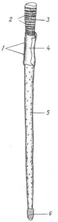
Fig. 158. Diagram of vestimentiferan body Structure:
1 — ciliated band; 2 — tentacular lamellae; 3 — obturaculum; 4 — vestimentum; 5, 6 — trunk and caudal regions
blood from the Blood vessels of the branchial filaments. The tentacular crown has a bright red color due to blood visible through the integument.
The second region features two wing-like body folds with highly developed musculature that wrap around the DORSAL SIDE OF the animal like a cloak, hence the name vestimentum (from Lat. *vestimentum* — garment, cloak). A broad ciliated band runs along the ventral side of this second region. Frenula are absent.
The third, trunk region — which accounts for half, and in large Riftia up to 80% of the body length — is not divided into two parts unlike that of frenulates. Similar to the outer surface of the wing-like folds of the second region, its integument is covered with wart-like glands that presumably secrete the tube material.
The fourth, caudal region, 2–3 cm long, consists of several dozen segments separated from one another by partitions. Each segment bears a girdle of small hooked setae.
The bulk of the trunk region is occupied by the trophosome — a nutritive organ consisting of numerous tubules intertwined with blood vessels and entirely packed with symbiotic autotrophic Bacteria that oxidize hydrogen sulfide or methane. The energy released in this process is used for the METABOLISM/21.html">Chemosynthesis of organic matter from carbon dioxide in the presence of highly active Enzymes. The blood continuously delivers oxygen and hydrogen sulfide (H2S) to the bacterial symbionts, while receiving Organic compounds synthesized by the bacteria, which are required to nourish the Tissues and Organs of the vestimentiferan. Studies of small pogonophorans of the class Frenulata, particularly the genera Siboglinum and Oligobrachia inhabiting the ordinary sea floor, have demonstrated that they too harbor symbiotic bacteria in the post-annular part of the trunk, filling the Cells of the coelomic epithelium between blood vessels. These animals also derive a significant portion of their nutrients through the chemosynthesis of their symbionts.
The embryonic development of afrenulates remains unstudied; it is only known that eggs are not released into the tube. Observations of Riftia pachyptila in aquaria have shown that its eggs are small but yolk-rich. Because the eggs are buoyant, they slowly float to the surface. This suggests that the eggs and larvae of afrenulates can be dispersed by currents, which is vital for these animals when the hydrothermal vent they inhabit becomes extinct — an event that occurs within no more than 10 years. A newly settled juvenile (Ridgeia sp., Oasisia alvinae, Riftia pachyptila) is 0.15 mm long and immediately begins secreting a tube, initially flask-shaped and subsequently elongating. At a length of 0.25–0.30 mm, the vestimentiferan possesses a pair of well-developed tentacles (Fig. 159), and at 5–6 mm it is already capable of withdrawing into the tube and closing its opening with the operculum.
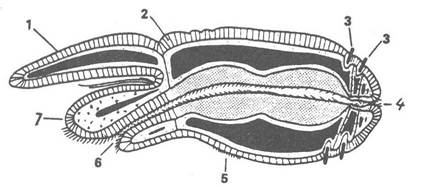
Fig. 159. Diagram of a juvenile vestimentiferan at the two-tentacle stage:
1 — tentacle; 2 — epidermis; 3 — setae; 4 — anus; 5 — ventral ciliated band; 6 — Mouth; 7 — cephalic process
At early Selection/3.html">Stages of development, vestimentiferans possess a Digestive System: the mouth leads to a funnel-shaped Pharynx, followed by a ciliated Esophagus that passes through the Brain and continues into the midgut, the latter transitioning into a voluminous hindgut. It is the midgut that transforms into the trophosome. Juveniles ingest free-living sulfur bacteria, which abound near sulfur vents, through the mouth. Once inside the midgut, these bacteria are engulfed by phagocytes; rather than digesting them, the phagocytes transport the bacteria into the intestinal folds where they settle, and the sulfur bacteria, now functioning as symbionts, begin to multiply. The digestive system Functions for quite a long time — until The formation of several hundred tentacles — supplying the bacteria with oxygen and hydrogen sulfide or methane. Subsequently, the supply function shifts to the Circulatory system, and the gut gradually degenerates. First, the midgut separates from the hindgut, which regresses and disappears along with the anus. Later, the connection with the esophagus is lost; the esophagus degrades, though persisting in adults as a thin strand passing through the brain. Research on vestimentiferans is only just beginning.
Last update: 13/08/2026
Editorial and Educational Adaptation: This material has been compiled based on the primary/original source text. The project team performed an editorial review, corrected technical inaccuracies, structured sections, and adapted the content for an educational format.
What was processed:
- elimination of formatting defects (OCR errors, structural breaks, corrupted characters);
- editorial organization of content;
- standardization of terminology in accordance with academic sources;
- verification of factual statements against the original source text.
All mentions of the author, publication year, and origin of the primary text have been preserved in accordance with the source.