INVERTEBRATE ZOOLOGY IN THREE VOLUMES - BOOK 3 - G.Y. Shcherbak - 1997
PHYLUM POGONOPHORA
CLASS FRENULATA
Most described species of pogonophores belong to this Class, found predominantly at depths exceeding 3,000 m at temperatures of -3 to +13 °C and normal Water salinity. In northern seas, pogonophores have also been found in shallow waters. Some species, such as Siboglinum caulleryi, occur at depths ranging from 20 to 8,100 m.
The size of frenulates ranges from 5 to 36 cm with a diameter of 0.1–2.5 mm.
The body consists of four regions (Fig. 150). The anterior and shortest body region bears a relatively long tentacular apparatus comprising from one to several hundred tentacles. In all pogonophores, the tentacles are body outgrowths containing a coelomic channel through which afferent and efferent Blood Vessels pass. Two rows of ciliated Cells extend along the tentacles. The tentacles bear rows of numerous, very delicate and thin pinnules; in frenulates, a pinnule is an overgrown epithelial Cell. Two blood capillaries enter each pinnule.
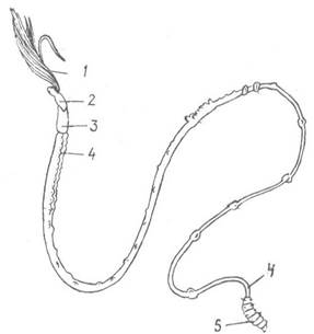
Fig. 150. A pogonophore from the family Polybrachiidae, without its tube:
1 — tentacles; 2, 3, 4, 5 — first, second, third, and fourth body regions, respectively
The tentacles are arranged in a circle or horseshoe shape. In many species, they lie close to one another without fusing, whereas in Representatives of the genera Lamellisabella and Spirobrachia, they unite into tentacular plates, forming a tentacular cylinder in the former and a spirally coiled tube in the latter (Fig. 151).
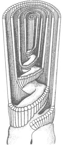
Fig. 151. Cross-section through the tentacular plates of Lamellisabella zachsi
The second, somewhat longer region bears two cuticular ridges called the frenulum; these rest against the edges of the tube and serve as a support for the anterior body region (Fig. 152).
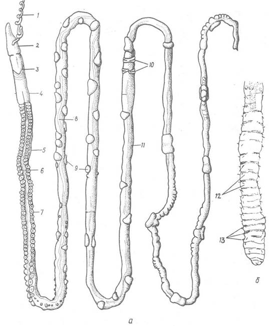
Fig. 152. Female of Siboglinum caulleryi without tube (a) and detached posterior end of the body (b):
1 — tentacle (partially cut off); 2 — first body region; 3 — frenulum; 4 — second body region; 5 — preannular part of the trunk region; 6 — ciliated band; 7 — attachment papillae with chitinous plates; 8 — blood vessel; 9 — glandular papillae; 10 — girdles; 11 — postannular part of the body; 12 — pseudosegments; 13 — setae
The third, trunk region is the longest. It is divided into two parts: the anterior preannular part, which extends to special girdles consisting of two to three rows of denticulate setae, and the posterior postannular part, located behind these girdles. A broad ciliated band is situated on the ventral side of the preannular part.
Numerous rounded or pear-shaped attachment papillae are located on the trunk (with the exception of species of the genus Sclerolinum, which lack papillae). In some species, the papillae are randomly scattered in large numbers over the entire trunk, whereas in others they are much fewer and located either at the anterior end of the body or metamerically distributed over the entire body. Small chitinoid plates are often present on the papillae. The papillae and denticulate setae serve to anchor the body against the inner wall of the tube.
The fourth region, which terminates the pogonophore body, is short like the first one and exhibits external segmentation; as a rule, it bears metamerically arranged setae that participate in the burrowing Movements of the pogonophore.
The tubes inhabited by frenulates vary in Structure (Fig. 153), but the anterior end of the tube is always wider than the posterior one. The first body region with tentacles protrudes outward through its opening, whereas only the fourth body region protrudes through the narrow posterior opening of the tube, performing peristaltic contractions to function as a burrowing organ. This region easily breaks off together with the posterior part of the tube it inhabits and therefore remained unknown to researchers for a long time, as it remained embedded in the substrate when the animal was pulled out.
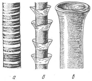
Fig. 153. Pogonophore tubes:
a — Siboglinum fedotovi; b — Polybrachia annulata; c — aperture of the tube of Spirobrachia beklemischevi
Various sessile invertebrates — Sponges, hydrozoan polyps, bryozoans, and others — settle on the tubes of many species. The pattern of distribution of these organisms indicates that a significant portion of the tube is embedded in the sediment. Throughout life, the posterior end of the tube is extended, causing it to sink deeper and deeper into the substrate.
Each body region has its own coelomic compartment (Fig. 154): in the first region, the coelom is unpaired, sac- or horseshoe-shaped, with its branches extending into all tentacles, whereas the second through fourth regions contain paired coelomic sacs.

Fig. 154. Diagram of the Internal Structure of the anterior part of the body in frenulate pogonophorans:
a — dorsal view; b — left view; 1, 2 — coelomic canals and Blood vessels of the tentacles; 3 — coelom of the first body segment; 4 — coelomoduct; 5, 6 — second body segment and its coelom; 7 — coelom of the third body segment; 8, 9 — ventral and dorsal blood vessels; 10 — main blood vessel; 11 — Brain; 12 — lateral blood vessel; 13 — nerve trunk; 14 — vas deferens; 15 — Origin of the tentacular nerves; 16 — Pericardium; 17 — Heart; 18 — male genital opening
The Digestive System is completely absent. The question of how pogonophorans feed was debated for a long time. It is now established that, like some other marine invertebrates (Molluscs, polychaetes, Echinoderms, etc.), pogonophorans absorb dissolved organic matter from the water, in particular Amino Acids, which penetrate their bodies through the body surface, including the tentacles. The body wall acts as a selective filter that allows small molecules to pass through while retaining larger ones. In small species, the Uptake of Amino acids accounts for a significant portion of their total METABOLISM. Furthermore, a second unique feeding mechanism involving Chemosynthesis products was discovered first in representatives of the class Afrenulata and later in some frenulates, owing to their Symbiosis with sulfur-oxidizing Bacteria (see p. 201).
The excretory system is represented by long ciliated canals—coelomoducts—connecting the coelom of the first body segment to the external environment.
The Circulatory system is closed. Blood flows through the dorsal vessel toward the anterior end of the body; near the Base of the tentacles, this vessel expands into a muscular heart whose contractions pump blood into the afferent Vessels of the tentacles, where most of the gas exchange takes place.
Then, via efferent vessels, the blood enters the ventral vessel and flows backward. In the posterior part of the trunk region, the dorsal and ventral vessels are connected by numerous transverse vessels. Pogonophoran blood is red and contains Hemoglobin.
The Gonads in both males and females are located in the trunk region of the body. Females have a pair of Ovaries in the anterior part of this region, from which mature egg cells enter the coelomic fluid via rupture of the Ovary wall and are subsequently expelled from the body through specialized oviducts. Males possess two long Seminal Vesicles in the posterior half of the same region, from which two vasa deferentia extend toward the anterior end of the trunk, opening on the DORSAL SIDE OF the body. Numerous flattened or spindle-shaped spermatophores with a long thread are formed within the vasa deferentia. It is likely that spermatophores actively move from males to females.
The female deposits eggs into the anterior portion of her own tube, where Fertilization and the entire process of embryonic development take place. Cleavage is total, unequal, and spiral, and asynchronous in the early stages. Gastrulation occurs via delamination and epiboly in some species (Siboglinum) and via invagination in others (Oligobrachia). The gastrula temporarily develops an intestinal rudiment, from which a pair of
coelomic pouches separates (enterocoely), and a blastopore is also present. Subsequently, the blastopore disappears, the endodermal gut rudiment disintegrates and is utilized by the embryo as nutritive material, while only the coelomic pouches persist. The embryo gradually elongates and segments; the fourth (posterior) segment with its coeloms is the first to separate, followed by the third segment, while The formation of tentacles begins at the anterior end. Finally, the anterior end divides into the First and Second segments. The newly hatched larva possesses two ciliated bands (Fig. 155), after which the cilia disappear, and the larval body grows in length, particularly its third segment.
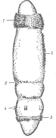
Fig. 155. Larva of Siboglinum fiordicum:
1 — anterior ciliated band; 2 — ventral ciliated strip; 3 — posterior ciliated band; 4 — setae; 5 — annular furrow between the second and third body segments
The class Frenulata is divided into two orders.
Order Athecanephria. This order includes frenulates in which the coelom of the first body segment is sac-like; The Heart is surrounded by a pericardium, and the coelomoducts are spaced apart from each other. Tentacles are few in number. This order includes one of the most speciose genera of pogonophorans, Siboglinum, representatives of which possess only a single tentacle. Two tentacles lacking pinnules are characteristic of the aforementioned species of the genus Sclerolinum, which bore holes in sunken pieces of wood and inhabit them (Fig. 156).
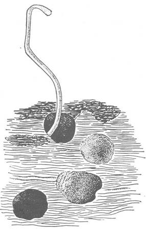
Fig. 156. Tube of Sclerolinum sp. embedded in a piece of wood
Order Thecanephria. In representatives of this order, the anterior coelom is horseshoe- or spiral-shaped. The coelomoducts are positioned close together in the middle of the first coelom, and a pericardium is absent. Of the three families belonging to this order, the Polybrachiidae possess free tentacles and a membranous anterior portion of the tube, whereas in members of the other two families, the tentacles are fused into a complex tentacular apparatus, and the tube opening is funnel-shaped. The number of tentacles is significantly greater than in representatives of the first order. For instance, Lamellisabella minuta has ten tentacles, whereas one of the largest frenulate pogonophorans, Galathealinum arcticum, has up to 268.
Last update: 13/08/2026
Editorial and Educational Adaptation: This material has been compiled based on the primary/original source text. The project team performed an editorial review, corrected technical inaccuracies, structured sections, and adapted the content for an educational format.
What was processed:
- elimination of formatting defects (OCR errors, structural breaks, corrupted characters);
- editorial organization of content;
- standardization of terminology in accordance with academic sources;
- verification of factual statements against the original source text.
All mentions of the author, publication year, and origin of the primary text have been preserved in accordance with the source.