INVERTEBRATE ZOOLOGY IN THREE VOLUMES - BOOK 3 - H.I. Shcherbak - 1997
DEUTEROSTOMIA
Deuterostomes are a major group of animal phyla sharing common features in Structure and embryonic development that sharply distinguish them from the protostome phyla discussed previously. The deuterostomes comprise three primary phyla: Hemichordata (hemichordates), Echinodermata (Echinoderms), and Chordata (Chordates). Some researchers also include the phyla Pogonophora (pogonophorans) and Chaetognatha (arrow worms) in this group, though this Classification remains subject to considerable debate.
Deuterostomes are characterized by a distinct suite of features, most of which pertain to Embryogenesis.
Cleavage of the egg in deuterostomes is radial and typically indeterminate. This cleavage yields a blastula, which undergoes invagination to form a two-layered gastrula. Unlike in most invertebrates, the blastopore does not develop into the adult Mouth. Instead, the anus forms at the site of the blastopore, while the definitive (secondary) mouth opens on the ventral side at the opposite (anterior) end of the body (The phenomenon of deuterostomy, Fig. 160).
Class="center">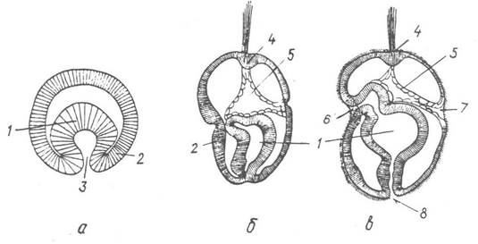
Fig. 160. KEY FEATURES OF deuterostome embryonic development:
a — gastrula; b–c — successive stages of secondary mouth formation in Balanoglossus (phylum Hemichordata); 1 — gut rudiment; 2 — ectoderm; 3 — blastopore; 4 — apical plate; 5 — coelom I; 6 — secondary mouth; 7 — coelomic pore; 8 — anus
The third germ layer (mesoderm) arises via lateral outpocketings of the entodermal gut, known as enterocoelous pouching (Fig. 161). These outpocketings initially appear as three pairs of lateral pouches (the first pair may be unpaired), which subsequently pinch off from the gut and position themselves on either side of it, developing into coelomic sacs whose walls give rise to the mesodermal structures of the adult animal.

Fig. 161. Formation of the coelom in Saccoglossus kowalevskii:
1 — apical plate; 2, 3, 4 — coeloms I, II, and III, respectively; 5 — gut endoderm
Consequently, the coelom in deuterostomes is tripartite (trimeric); it consists of three pairs of coelomic sacs (the first of which may be fused), with the left-side coeloms typically more developed than the right. Coelomic trimerism is most pronounced in hemichordates; in chordates, it is primarily the last (third) pair of coeloms that undergoes major development; whereas in echinoderms, the coeloms experience the most radical reorganization, retaining their tripartite arrangement only in the larval stage, while in adults they give rise to various Organ Systems.
The characteristic larva of deuterostomes is the dipleurula — a bilaterally symmetrical planktonic larva possessing a complete gut with a secondary mouth, three pairs of coelomic sacs, an apical plate with a Brain rudiment, and a sinuous ciliated band encircling the body (see p. 210). The term dipleurula means "double-sided" larva, highlighting its inherent bilateral Symmetry. While typical of hemichordates and echinoderms, the dipleurula larva is absent in chordates due to the embryonization of their development.
The integument of deuterostomes consists of a single-layered ectodermal epidermis and a mesodermal dermis (cutis).
The Skeleton, when present, is internal, cartilaginous or calcareous, and of mesodermal origin.
The Nervous system in deuterostomes typically takes the form of epithelial strips that in many cases sink beneath the epithelium to form nerve tubes; alternatively, a diffuse subepidermal nerve plexus may be retained. The brain, when present, is secondary; it forms as an expansion of the anterior end of the nerve tube rather than originating from the larval apical plate.
Last update: 13/08/2026
Editorial and Educational Adaptation: This material has been compiled based on the primary/original source text. The project team performed an editorial review, corrected technical inaccuracies, structured sections, and adapted the content for an educational format.
What was processed:
- elimination of formatting defects (OCR errors, structural breaks, corrupted characters);
- editorial organization of content;
- standardization of terminology in accordance with academic sources;
- verification of factual statements against the original source text.
All mentions of the author, publication year, and origin of the primary text have been preserved in accordance with the source.