IMMUNOLOGY - Roit I. - Mir 2000
Chapter 6. Antibodies and Cellular Receptors for Them
CELLULAR RECEPTORS FOR ANTIBODIES
There are Three types of Cell surface receptors for IgG
Cellular receptors for IgG mediate a range of antibody effector Functions. Cross-linking of receptor-bound Ig Antibodies by an antigen initiates various biological activities of The Cell, with different receptors capable of inducing identical activities, among which the primary ones are phagocytosis, antibody-dependent cellular cytotoxicity, mediator release, and antigen presentation.
To date, three groups of human IgG receptors have been identified on the cell surface: FcγRI (CD64), FcγRII (CD32), and FcγRIII (CD16). All of them possess extracellular domains that are largely homologous to the V regions of IMMUNOGLOBULINS, meaning they belong to the immunoglobulin superfamily of molecules, much like the IgA-specific receptor FcαR.
Properties and distribution of receptors for IgG. The FcγRI (CD64) receptor on human Cells binds monomeric IgG with high affinity (108—109 M-1) and has a more restricted distribution than other receptors.
The FcγRII (CD32) receptor is expressed on A wide variety of cells, often serving as the sole Ig receptor. It binds IgG with low affinity (<107 M-1), and only when the latter is incorporated into immune complexes or aggregates.
The FcγRIII (CD16) receptor is heavily glycosylated and is expressed in isoforms with molecular weights ranging from 50 to 80 kDa. The FcγRIIIa form is expressed by macrophages, NK cells, and certain T cells; this form binds monomeric IgG as well as IgG incorporated into immune complexes with an affinity of 3 x 10⁷ M-1. In the FcγRIIIb form, which is anchored to The cell membrane via GPI (phosphatidylinositol glycan), the receptor is expressed exclusively by granulocytes; FcγRIIIb binds IgG with low affinity (<107 M-1).
Additional diversity of cellular receptors for IgG. The three types of Fcγ receptors occur in 12 different isoforms, and genetic polymorphism has also been described for FcγRII and FcγRIII. Alongside this intrinsic heterogeneity, another type of diversity has been established, driven by the expression of these receptors on the cell surface as complexes with polypeptide chains of different origin. Two Types of such chains have been identified, associating with various receptor types:
✵ FcγRI, similarly to FcεRI, forms a complex with a disulfide-linked γ-chain dimer,
✵ FcγRIIIa can associate with either the same γ-chain dimers, ζ-chain dimers (from the TCR complex), or heterodimers of γ- and ζ-chains.
The associated chains presumably facilitate receptor expression on The Plasma Membrane and, furthermore, appear crucial for intracellular signal Transduction. In the case of FcγIIIb, which is membrane-anchored via GPI, neither γ- nor ζ-chains seem necessary (Fig. 6.19).
Class="center">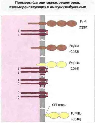
Fig. 6.19. Structure of human Fcγ receptors: FcγRI (expressed by monocytes), FcγRIIa (expressed by monocytes and neutrophils), FcγRIIIa (expressed by monocytes and surface-bound like a conventional transmembrane protein), and FcγRIIIb (expressed by neutrophils and anchored to the cell membrane via phosphatidylinositol glycan [GPI]). Each receptor type belongs to the immunoglobulin superfamily of molecules and features two or three extracellular immunoglobulin-like domains. Some receptors are now known to be complexes of distinct disulfide-linked subunits. FcγRI and FcγRIII receptors are associated with a γ-chain dimer initially described as part of the high-affinity FcεRI complex (see Fig. 6.20). The FcγRIIIa receptor is associated with a ζ-chain dimer previously identified within the TCR-CD3 complex. FcγRIIIa may associate with either homodimeric (γ-γ or ζ-ζ) or heterodimeric (γ-ζ) subunits, which appear essential for surface expression and Transmembrane Signal Transduction. Upon interacting with an immunoglobulin, FcγRI likely binds to the structural region surrounding the leucine residue at position 235 of the CH2 domain. Such a structural motif is present in the heavy chains of IgG1, IgG3, and IgG4.
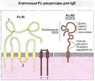
Fig. 6.20. According to this model, FcεRI consists of four components: a single α-chain with two disulfide-bonded loops resembling immunoglobulin domains, a single β-chain with two extracellular segments, and a pair of nearby disulfide-linked γ-chains (red bar). The α-chain plays a crucial role in binding FcεRI to IgE. The hypothetical model of FcεRII is based on sequencing data and Homology with animal Lectins. Proteolysis of these receptors yields several types of IgE-binding factors, including soluble CD23 molecules (25 kDa molecular weight) containing a lectin domain. Judging by the pronounced upregulation of FcεRII expression on lymphocytes cultured in the presence of IgE, this immunoglobulin inhibits receptor proteolysis.
IgE binds to two distinct cellular Fcε receptors
Currently, two different cellular receptors for IgE are known (Fig. 6.20). Mast cells and basophils feature the high-affinity "classical" FcεRI receptor, which belongs to the immunoglobulin superfamily. A substantially distinct low-affinity Fc receptor for IgE has been identified on leukocytes, including lymphocytes. This low-affinity receptor does not belong to the immunoglobulin superfamily, but shows significant homology to certain animal lectins, such as the mannose-binding lectin.
FcεRI - high-affinity cellular receptor for IgE. The FcεRI molecule is formed by 4 polypeptide chains (see Fig. 6.20). Its glycosylated α-chain (45 kDa) is exposed on the cell surface. Antibodies against the α-chain can block IgE binding to this receptor and independently induce histamine release from rat leukemic basophils. The carbohydrate Components of the α-chain presumably protect it (as is the case with many other cell surface Proteins) from serum proteases, yet they are not essential for IgE binding or IgE-mediated histamine release.
A single β-chain (33 kDa) and two disulfide-linked γ-chains (9 kDa) are essential components of the αβγ2 receptor unit. They are required for cell surface receptor expression and presumably for transmembrane signal transduction.
The FcεRI receptor interacts with the C-terminal region of IgE heavy chains, specifically the Cε2 and/or Cε3 domains. This binding is highly specific and characterized by a very high affinity constant (~1010 M-1). However, neither receptor interaction with monomeric IgE nor the binding of a specific Ligand by a single IgE molecule appears to activate mast cells or basophils, as no histamine release occurs. Triggering degranulation requires multiple cell-surface-bound IgE molecules to be cross-linked by an antigen or other degranulation-inducing ligands.
Carbohydrate components of the IgE molecule are likely not involved in the interaction with FcεRI, though they are important for IgE secretion by B cells.
It was previously believed that high-affinity receptors for IgE existed exclusively on mast cells and basophils, but recent findings indicate they may also be present on Langerhans cells and stem cells.
FcεRII - low-affinity cellular receptor for IgE. The human lymphocyte FcεRII, or CD23 antigen (45 kDa), contains a transmembrane domain typical of a membrane-bound molecule, yet it is positioned unusually "upside down" in the membrane, i.e., with its C-terminus facing the extracellular space (Fig. 6.20). Unlike other Fc receptors, it belongs not to the immunoglobulin superfamily, but to the phylogenetically older animal lectin superfamily.
To date, two human FcεRII forms have been identified, cloned, and sequenced; they share identical extracellular domain structures but differ in their N-terminal cytoplasmic region. The FcεRIIa receptor is constitutively expressed on normal B cells, whereas FcεRIIb expression is induced by the cytokine IL-4 on T cells, B cells, monocytes, and eosinophils. This expression is often upregulated on B cells and monocytes in eczema patients, and on lymphocytes in hay fever.
Last update: 13/08/2026
Editorial and Educational Adaptation: This material has been compiled based on the primary/original source text. The project team performed an editorial review, corrected technical inaccuracies, structured sections, and adapted the content for an educational format.
What was processed:
- elimination of formatting defects (OCR errors, structural breaks, corrupted characters);
- editorial organization of content;
- standardization of terminology in accordance with academic sources;
- verification of factual statements against the original source text.
All mentions of the author, publication year, and origin of the primary text have been preserved in accordance with the source.