MICROBIOLOGY - M.H. Serhiichuk - 2008
Chapter 5. MICROBIAL METABOLISM
MECHANISMS OF CARBON ASSIMILATION IN MICROORGANISMS
Carbon sources for microorganisms include both organic and inorganic substances (carbon dioxide, carbonates).
Assimilation of inorganic carbon. Carbon dioxide is the most abundant form of carbon on Earth. Many microorganisms, known as autotrophs, have The ability to incorporate CO2 into cellular carbon. This process is referred to as CO2 fixation.
Carbon dioxide fixation. Microorganisms utilize four distinct pathways for carbon dioxide fixation:
1. The Calvin Cycle (reductive Pentose Phosphate Pathway or ribulose biphosphate pathway, RuBP);
2. The reductive Tricarboxylic Acid Cycle (Evans-Buchanan-Arnon cycle, rTCA);
3. The reductive acetyl-CoA pathway (Wood-Ljungdahl pathway, rACA);
4. The 3-hydroxypropionate cycle.
The Calvin cycle (Fig. 5.30) is The most significant biosynthetic pathway on Earth. It is utilized by the majority of photosynthetic organisms, including plants, cyanobacteria, purple Bacteria, and green bacteria. The Calvin cycle consists of three phases:
1. Carbon dioxide fixation. CO2 is coupled with ribulose-1,5-bisphosphate, a five-carbon compound, yielding two molecules of 3-phosphoglyceric acid. One of the two carboxyl groups in this compound originates from CO2. The reaction is catalyzed by ribulose-1,5-bisphosphate carboxylase-oxygenase (RuBisCo), the signature key enzyme of this cycle.
Class="center">Ribulose-1,5-bisphosphate + CO2 + H2O → 3-Phosphoglyceric acid (2 molecules)
2. Reduction of phosphoglyceric acid carboxyl groups to aldehydes. The carboxyl group of 3-phosphoglyceric acid is first activated via phosphorylation to form 1,3-diphosphoglyceric acid, which is subsequently reduced to 3-phosphoglyceraldehyde. This stage consumes virtually all the energy and reducing equivalents required for CO2 fixation. A portion of the 3-phosphoglyceraldehyde is converted into fructose-6-phosphate to fuel biosynthetic processes.
3-Phosphoglyceric acid + ATP → 1,3-Diphosphoglyceric acid + ADP
1,3-Diphosphoglyceric acid + NADPH2 → 3-Phosphoglyceraldehyde + NADP+ + Pi
3. Regeneration of the CO2 acceptor. The remaining Reactions of the cycle regenerate ribulose-1,5-bisphosphate. These begin with the synthesis of fructose-6-phosphate from two triose phosphate molecules. This is followed by a rearrangement of the carbon Skeleton to generate pentose-5-phosphates, mirroring the reverse (reductive) reactions of The pentose phosphate pathway. The regeneration of the CO2 acceptor is completed by the ATP-dependent phosphorylation of ribose-5-phosphate to ribulose-1,5-bisphosphate. This process is catalyzed by the second specific key enzyme of the Calvin cycle, phosphoribulokinase.
Energy balance of the Calvin cycle. To generate a single net molecule of fructose-6-phosphate—which can then be withdrawn from the cycle for Biosynthesis—six molecules of ribulose-1,5-bisphosphate must be carboxylated, meaning that 6CO2 are fixed. Following carboxylation, these six molecules of ribulose-1,5-bisphosphate are converted into 12 molecules of 3-phosphoglyceric acid, then into 12 molecules of 1,3-diphosphoglyceric acid, and finally reduced to 12 molecules of 3-phosphoglyceraldehyde. The Cell expends 12 ATP and 12 NADPH2 on these transformations. An additional 6 ATP are consumed during the regeneration of the CO2 acceptor to phosphorylate ribose-5-phosphate into ribulose-1,5-bisphosphate. Thus, the synthesis of a single glucose molecule "costs" the cell 18 ATP and 12 NADPH2. The overall stoichiometric equation for CO2 fixation via the Calvin cycle is as follows:
6CO2 + 12 NADPH2 + 18 ATP → C6H12O6 + 12 NADP+ + 18 ADP + 18 Pi
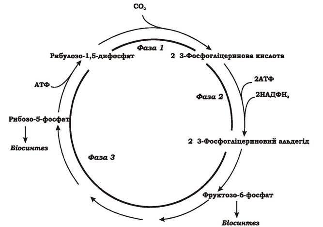
Fig. 5.30. The Calvin cycle
(reductive pentose phosphate pathway or ribulose bisphosphate pathway)
The reductive tricarboxylic acid cycle was described by Evans, Buchanan, and Arnon in 1966 in the green sulfur bacterium Chlorobium thiosulfatophilum. It was later discovered in certain sulfate-reducing bacteria and thermophilic hydrogen-oxidizing bacteria of the genus Hydrogenobacter. The reductive TCA cycle is essentially a modified Krebs cycle operating in reverse, in a reductive direction (Fig. 5.31).
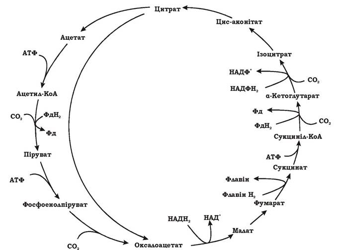
Fig. 5.31. Reductive tricarboxylic acid cycle (synonyms: Evans-Buchanan-Arnon cycle, reductive TCA cycle)
There are full and short reductive TCA cycles. As a result of one full turn of the cycle, four molecules of СО2 are fixed and OAA is synthesized. Since OAA itself is an intermediate product of the cycle, after one rotation, one molecule of OAA is regenerated, while a second one is synthesized from four fixed and reduced СО2 molecules. During a short cycle, two fixed СО2 molecules yield one molecule of acetate.
As can be seen from the figure, the full and short cycles share the same sequence of reactions from OAA to citrate. That is, starting from OAA, a complete Rotation of the short reductive TCA cycle leads to OAA regeneration and the synthesis of acetate from СО2. Ultimately, it is the short reductive TCA cycle—which produces acetyl-CoA from 2СО2—that is the reverse of the Krebs cycle. The full reductive TCA cycle involves four СО2 fixation and reduction reactions:
1. Acetyl-CoA + СО2 + Ferredoxinred → Pyruvate + CoA + Ferredoxinox
2. Succinyl-CoA + СО2 + Ferredoxinred → α-Ketoglutarate + CoA + Ferredoxinox
3. α-Ketoglutarate + СО2 + NADPH2 → Isocitrate + NADP+
4. Phosphoenolpyruvate + СО2 + → OAA + Pi
Reactions (2) and (3) occur in both the standard Krebs cycle and the reductive TCA cycle, whereas reactions (1) and (4) are unique to the reductive TCA cycle. Reactions (1) and (2) are driven by ferredoxins rather than NADPH2. СО2 assimilation requires Energy Expenditure. The Discovery of the Evans–Buchanan–Arnon cycle confirmed the evolutionary link between heterotrophic and photoautotrophic microorganisms.
The reductive acetyl-CoA pathway is found in the METABOLISM of autotrophic sulfate reducers, homoacetogens, and methanogens. Unlike other СО2 assimilation pathways, it is non-cyclic (Fig. 5.32). One molecule of СО2 is bound by a special cofactor, tetrahydrofolic acid, and reduced to a methyl group. A second СО2 molecule is reduced to a carbonyl (C=O) group by the enzyme carbon monoxide dehydrogenase. The methyl and carbonyl groups are then combined into acetyl-CoA, which is utilized in further biosynthetic processes. Two molecules of СО2 are reduced at the expense of 4Н2.
Overall equation of the process: 4Н2 + 2СО2 = Acetate + 2Н2О
The acetyl-CoA pathway can also operate in the reverse, oxidative direction. Heterotrophic sulfate reducers employ the oxidative acetyl-CoA pathway for acetate Cleavage (Fig. 6.26).
The 3-hydroxypropionate СО2 fixation cycle (Fig. 5.33) was discovered in green non-sulfur photosynthetic bacteria of the genus Chloroflexus and certain archaea.
A full turn of this cycle fixes 2СО2 and synthesizes glyoxylate (CHOCOOH). The first СО2 molecule is accepted by acetyl-CoA to form malonyl-CoA, while the second СО2 molecule combines with propionyl-CoA to yield methylmalonyl-CoA. The terminal intermediate of the cycle, malyl-CoA, is cleaved into acetyl-CoA and glyoxylate. Acetyl-CoA fixes СО2 anew in the next turn, whereas glyoxylate is channeled into biosynthesis. The process can be described by the following equation:
2НСО3- + 2 NADPH2 + 3Н+ + 3 ATP → CHO-COO- + 2 NADP+ + 3 ADP + 3 Pi
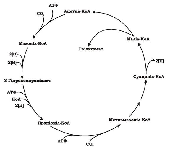
Fig. 5.32. Reductive acetyl-CoA pathway (synonym: Wood–Ljungdahl pathway): THF, tetrahydrofolic acid

Fig. 5.33. 3-Hydroxypropionate cycle
The electron Donors and The Nature of many Enzymes involved in this cycle remain unstudied.
Formaldehyde assimilation. Methylotrophic microorganisms utilize three pathways for formaldehyde (СН2О) assimilation:
1. Serine pathway.
2. Ribulose monophosphate cycle (hexulose phosphate pathway, RuMP).
3. Dihydroxyacetone pathway.
The first two pathways are typical of methylotrophic bacteria, whereas the last one is restricted to methylotrophic Yeasts.
The serine pathway is a cyclic process whose end product is acetyl-CoA, synthesized from one molecule of formaldehyde and one molecule of СО2 (Fig. 5.34).
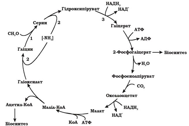
Fig. 5.34. The serine pathway of formaldehyde assimilation
The first reaction of this cycle involves the interaction of formaldehyde (C1) with Glycine (C2) to form serine (C3). This reaction is catalyzed by hydroxymethyltransferase, which requires tetrahydrofolic acid (THF) as a cofactor. The process proceeds via THF-bound intermediates. When formaldehyde attaches to the cofactor of hydroxymethyltransferase, 5,10-methylenetetrahydrofolic acid (methylene-
THF) is formed. Through reaction (1) (Fig. 5.34, 1), formaldehyde is transferred from methylene-THF to glycine, yielding serine.
In the subsequent reactions (Fig. 5.34, 2), an amino group is transferred from serine to glyoxylate. As a result, serine is converted into hydroxypyruvate, while glyoxylate is converted into glycine. Glycine then reacts with another formaldehyde molecule, and hydroxypyruvate undergoes further transformation (Fig. 5.34, 3).
Hydroxypyruvate is reduced to glyceric acid, which is subsequently phosphorylated to 2-phosphoglycerate with the consumption of 1 ATP. At this point, the serine pathway branches: a portion of the 2-phosphoglyceric acid is converted into 3-phosphoglyceric acid for biosynthesis, while the remainder is transformed into phosphoenolpyruvate (PEP). PEP (C3) is carboxylated with CO2 to form oxaloacetate (C4), which is reduced to malate and converted into malyl-CoA using ATP. Catalyzed by malyl-CoA lyase, malyl-CoA is cleaved into glyoxylate (a C2 compound), which re-enters the cycle, and acetyl-CoA (a C2 compound), which is channeled into biosynthesis.
The ribulose monophosphate cycle (Fig. 5.35) is a cyclic process of formaldehyde assimilation that consists of three stages:
1. Fixation. Ribulose-5-phosphate serves as the formaldehyde acceptor. Formaldehyde and ribulose-5-phosphate condense in a reaction catalyzed by hexulose phosphate synthase to yield hexulose-6-phosphate, which is then converted into fructose-6-phosphate by the action of hexulose phosphate isomerase. Hexulose phosphate synthase and hexulose phosphate isomerase are the only unique enzymes specific to this pathway. The remaining steps are catalyzed by Enzymes of the Entner-Doudoroff Pathway.
2. Cleavage. Fructose-6-phosphate is cleaved into C3 compounds via one of two routes:
a) fructose-6-phosphate is phosphorylated to fructose-1,6-diphosphate and subsequently cleaved into dihydroxyacetone phosphate and phosphoglyceraldehyde;
b) fructose-6-phosphate is first isomerized to glucose-6-phosphate, which is converted into D-glucono-1,5-lactone-6-phosphate, then into 6-phosphogluconate, and finally into 2-keto-3-deoxy-6-phosphogluconate (KDPG). KDPG is cleaved into 3-phosphoglyceraldehyde and pyruvate by KDPG aldolase.
One of the phosphotrioses (dihydroxyacetone phosphate or pyruvate) is directed toward biosynthesis.
3. Rearrangement. The final stage of the process is the rearrangement of phosphotrioses, which regenerates the formaldehyde acceptor, ribulose-5-phosphate. This can occur via several pathways, but overall, two molecules of xylulose-5-phosphate and one molecule of ribose-5-phosphate are converted into three molecules of ribulose-5-phosphate, completing the cycle.
Thus, as a result of the ribulose monophosphate cycle, three molecules of formaldehyde yield a triose (dihydroxyacetone phosphate or pyruvate), which is channeled into biosynthesis.
The overall equation of the process:
3CH2O + 2 ATP → Phosphotriose + 2 ADP + Pi
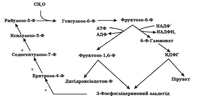
Fig. 5.35. Scheme of the ribulose monophosphate cycle for formaldehyde assimilation (synonym: hexulose phosphate pathway, RuMP)
* - reactions that can occur only when at least three molecules of formaldehyde are incorporated into the process
Assimilation of C2 substrates by microorganisms. When growing on media containing acetate as the sole carbon source, aerobic microorganisms assimilate acetyl-CoA via The Glyoxylate cycle.
The glyoxylate cycle is a sequence of reactions that converts two acetyl units into one C3 compound with the decarboxylation of the fourth carbon atom: 2C2 → 1C3 + 1C1 (Fig. 5.36).
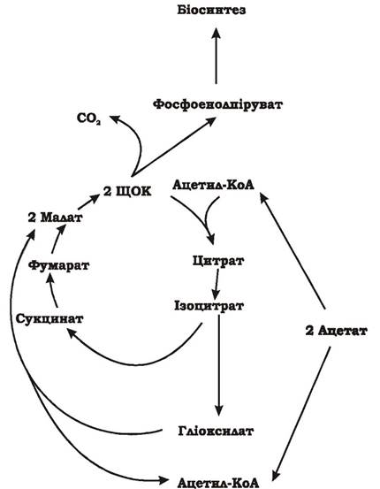
Fig. 5.36. The glyoxylate cycle of acetyl-CoA assimilation in microorganisms
In the first step of this cycle, acetyl-CoA condenses with oxaloacetate to form citrate, which is subsequently converted into isocitrate. Isocitrate is then cleaved by isocitrate lyase into two intermediates: glyoxylate and succinate.
Isocitrate → Glyoxylate + Succinate
In the second stage, two molecules of malate are synthesized. One molecule is formed directly from succinate via fumarate, while the second is produced through the Condensation of the glyoxylate generated in the first step with a second molecule of acetyl-CoA. This reaction is catalyzed by malate synthase:
Glyoxylate + Acetyl-CoA + H2O → Malate + CoA-SH
In the Third Stage, two molecules of malate yield two molecules of OAA, one of which is channeled into biosynthesis while the other Functions within the glyoxylate cycle. To enter biosynthetic pathways, OAA is decarboxylated to pyruvate or phosphoenolpyruvate.
Gluconeogenesis is the metabolic pathway through which glucose is synthesized from pyruvate (Fig. 5.37). While it shares many steps with Glycolysis—the process by which glucose is oxidized to pyruvate—these reactions proceed in the reverse (reductive) direction.
However, gluconeogenesis and glycolysis also exhibit several key differences. Unlike glycolysis, which yields 2 ATP during The oxidation of glucose, the Synthesis of glucose via gluconeogenesis consumes 6 ATP.

Fig. 5.37. Pathway of gluconeogenesis
Glycolysis:
Glucose + 2 NAD+ + 2 ADP + 2 Pi → 2 Pyruvate + 2 NADH2 + 2 ATP + 2H2O
Gluconeogenesis:
2 Pyruvate + 2 NADH2 + 4 ATP + 2 GTP + 6H2O → Glucose + 4 ADP + 2 GDP + 6 Pi + 2 NAD+ + 2H+ (GTP and ATP are mutually interconvertible: GDP + ATP → GTP + ADP). While glycolysis converts phosphoenolpyruvate into pyruvate in a single step catalyzed by pyruvate kinase with the net production of 1 ATP, gluconeogenesis converts pyruvate back into phosphoenolpyruvate via a two-step process involving an OAA intermediate, consuming 1 ATP and 1 GTP and requiring two separate enzymes: pyruvate carboxylase and phosphoenolpyruvate carboxykinase.
Last update: 13/08/2026
Editorial and Educational Adaptation: This material has been compiled based on the primary/original source text. The project team performed an editorial review, corrected technical inaccuracies, structured sections, and adapted the content for an educational format.
What was processed:
- elimination of formatting defects (OCR errors, structural breaks, corrupted characters);
- editorial organization of content;
- standardization of terminology in accordance with academic sources;
- verification of factual statements against the original source text.
All mentions of the author, publication year, and origin of the primary text have been preserved in accordance with the source.