Biological Membranes - A. N. Ogurtsov 2012
Biomembrane Electrogenesis
Mechanisms of Action Potential Generation
Myelination of Neuronal Axons
An Action Potential can travel along an axon at a speed of about 1 m/s. However, this Nerve Impulse transmission velocity is insufficient to support the complex and coordinated Muscle activity of mammals during movement.
For instance, in humans, The Cell bodies of Neurons controlling leg Muscles are located in the Spinal Cord, and their axons reach up to 1 meter in length. The coordination of muscle contractions during walking, running, or jumping would be impossible if nerve impulses took around a second to travel from the spinal cord to the muscles.
To increase the propagation speed of nerve impulses by 10–100 times, neuronal axons are wrapped in a myelin sheath (Figure 134). As a result, in a typical motor neuron, an action potential covers a length of 1 m in 0.01 s (i.e., at a speed of 100 m/s).
In unmyelinated neurons, the impulse propagation speed is roughly proportional to the axon diameter, because the thicker the axon, the easier it is for ions to diffuse along it.
The human Brain consists of densely packed myelinated neurons. If neurons were unmyelinated, maintaining the same impulse transmission speed as in myelinated neurons would require their axon diameter to be 10,000 times larger, increasing their mass by the same factor. Consequently, during evolution, the vertebrate brain could never have formed without the myelination of neurons.
Class="center">
Figure 134 - Diagram of axon myelination
The myelin sheath is a lamellar Membrane Structure wrapped in multiple layers around the axon, synthesized by glial Cells (Schwann cells) (Figure 134). The glial Cells of the Central Nervous system are called oligodendrocytes.
The myelin sheath around an axon is formed by numerous glial cells. Each myelination segment produced by a single glial cell is separated from the adjacent segment by an unmyelinated region of the axonal membrane about 1 µm wide, known as a node of Ranvier or simply a node.
Therefore, the axonal membrane is in direct contact with the extracellular fluid only at the nodes of Ranvier. Furthermore, all voltage-gated Na+ channels and Na+/K+-ATPases in myelinated axons are localized exclusively at the nodes. As a result, the influx of sodium ions into the axon during an action potential can occur only at the nodes of Ranvier (Figure 135).
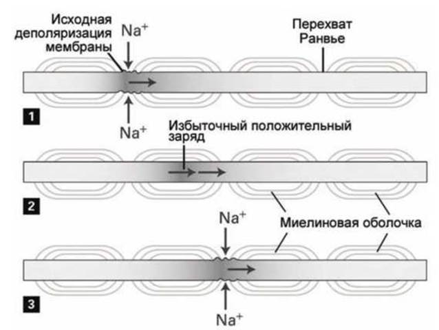
Figure 135 - Diagram of action potential propagation along a myelinated axon at successive time points 1, 2, 3
The excess cations generated by membrane depolarization at the node of Ranvier spread along the axon as a practically non-attenuating wave, driven by both the concentration gradient and the electrical potential gradient, toward the nearest node. Upon reaching the adjacent node, this wave of excess cations triggers membrane depolarization.
In effect, the propagation of a nerve impulse is a process of action potentials "skipping" from one node to the next. As a result, excitation is transmitted in a saltatory manner (saltatory conduction, from Latin saltare — to leap) from one node of Ranvier to another, at a higher speed and with a lower energy cost than in unmyelinated fibers of comparable diameter (Figure 136).
This is precisely why the propagation speed of action potentials in myelinated axons is tens to hundreds of times higher than in unmyelinated axons of the same diameter.
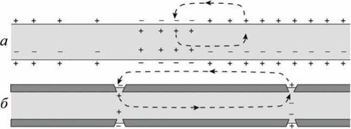
Figure 136 - Diagram of saltatory action potential propagation in a myelinated neuron: a - unmyelinated neuron; b - myelinated neuron
The significant (hundredfold) enrichment of membrane Ion Channels and pumps at the nodes is driven by the fact that the elements of the axonal Cytoskeleton and Extracellular matrix to which these Proteins attach are concentrated precisely at the nodes of Ranvier rather than in the myelinated Regions of the axonal membrane.
Last update: 13/08/2026
Editorial and Educational Adaptation: This material has been compiled based on the primary/original source text. The project team performed an editorial review, corrected technical inaccuracies, structured sections, and adapted the content for an educational format.
What was processed:
- elimination of formatting defects (OCR errors, structural breaks, corrupted characters);
- editorial organization of content;
- standardization of terminology in accordance with academic sources;
- verification of factual statements against the original source text.
All mentions of the author, publication year, and origin of the primary text have been preserved in accordance with the source.