Biological Membranes - A. N. Ogurtsov 2012
Electrogenesis of Biomembranes
Mechanisms of Action Potential Generation
Structure of Voltage-Gated Ion Channels
Studies on the molecular Structure of voltage-Gated Ion Channels have revealed a striking similarity in their overall architecture. Most of the investigated Voltage-Gated Potassium Channels exhibit a tetrameric structure composed of four identical subunits assembled into a functional complex that forms a central pore.
Each subunit of a voltage-gated potassium (K+) channel consists of six transmembrane a-helices, designated S1 through S6, along with a P-loop (Figure 130).
Class="center">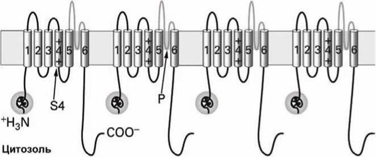
Figure 130 - Diagram of a voltage-gated potassium channel
The subunit fragment containing the S5 and S6 a-helices and the P-loop is structurally and functionally analogous to the subunit of a non-gated potassium channel, which is responsible for establishing and maintaining the resting Membrane Potential (Figure 91).
The S4 transmembrane a-helix contains numerous positively charged Lysine and Arginine residues and Functions as the voltage sensor of the membrane potential.
A globular cytoplasmic domain attached to the S1 a-helix near the N-terminus of the protein chain acts as a channel-inactivating (blocking) segment.
Voltage-gated sodium (Na+) and calcium (Ca2+) channels are monomeric Proteins consisting of four homologous domains (Figures 131 and 132).

Figure 131 - Diagram of a voltage-gated Na channel
Each of these domains is structurally and functionally similar to a single K+-channel subunit (Figure 130). However, unlike voltage-gated K+-channels, in which each subunit possesses its own channel-blocking segment (making a total of four in a functional K+-channel), monomeric voltage-gated channels contain only a single such blocking segment (H).
The selectivity filter of the channel is formed by four pore-forming loops (P-segments). Figure 132 illustrates the spatial arrangement of the four subunits of a Ca2+-channel, each comprising six transmembrane a-helices; the voltage-sensing a-helices (channel sensors) are shaded, and the pore-forming loops are oriented toward the interior of the channel pore.

Figure 132 - Schematic folding of structural elements in a voltage-gated calcium channel
Thus, it can be concluded that all ion channels are both structurally and functionally related and presumably evolved from a common ancestral protein that possessed six transmembrane a-helices.
The Role of voltage sensors in voltage-gated ion channels is played by four positively charged S4 a-helices. Their displacement triggers a conformational change in the channel protein that leads to the opening or closing of the ion channel (Figure 128).
The actual gate of the ion channel is formed by the N-termini of the S5 helices and the C-termini of the S6 helices from all four subunits.
Spontaneous inactivation of ion channels a few milliseconds after their opening is mediated by specific blocking segments resembling spherical, positively charged globules attached to the ends of unstructured protein tethers.
At rest, these globular domains at the N-termini of the four potassium channel subunits float freely in the Cytosol.
A few milliseconds after channel opening (following membrane depolarization), one of these globular heads moves through a lateral fenestration between two channel subunits and binds within a hydrophobic pocket in the central cavity of the channel, thereby blocking the flow of K+ ions (Figure 133).

Figure 133 - Diagram of voltage-gated potassium channel inactivation
Within a few milliseconds after membrane repolarization, the blocking segment is dislodged from the pore, and the protein returns to its resting state.
The ion channel is formed by four a-membrane domains, whereas the cytosolic ß-domains serve a regulatory function. The globular domains of the potassium channel are functionally equivalent to the blocking segment in the sodium channel.
Artificial Modification of the protein loops connecting the blocking globules to the channel body has demonstrated that shortening the connecting peptide tether accelerates channel inactivation, whereas lengthening the tether slows it down.
Last update: 13/08/2026
Editorial and Educational Adaptation: This material has been compiled based on the primary/original source text. The project team performed an editorial review, corrected technical inaccuracies, structured sections, and adapted the content for an educational format.
What was processed:
- elimination of formatting defects (OCR errors, structural breaks, corrupted characters);
- editorial organization of content;
- standardization of terminology in accordance with academic sources;
- verification of factual statements against the original source text.
All mentions of the author, publication year, and origin of the primary text have been preserved in accordance with the source.