Biological Membranes - A. N. Ogurtsov 2012
Structure and Functions of Biomembranes
Structure of Lipid Membranes
Model Lipid Membranes
Introduction/36.html">Biological Membranes generally have a highly complex Structure and exhibit relatively low resistance to mechanical, chemical, and electrical influences. Therefore, to study the fundamental PHYSICOCHEMICAL PROPERTIES OF Cell membranes, various artificial systems are widely used to model processes occurring in Biomembranes.
Artificial Membranes have a rather simple structure and high stability, which makes it possible to widely vary experimental conditions and obtain crucial insights into the potential mechanisms of biological membrane function.
Biological membranes can be described as complex multienzyme systems. Information about their structural Organization can be obtained by reconstituting them on artificial lipid membranes. The creation of artificial lipid membranes, which serve as physicochemical analogues of natural biological membranes, constitutes The Essence of the model approach aimed at elucidating the functional role of each individual component of a native biological membrane.
Three MAIN TYPES OF lipid models of biological membranes are most widely used:
1) lipid monolayers at the electrolyte solution–air interface;
2) planar bilayer lipid membranes;
3) Liposomes, which are closed vesicular structures consisting of one or more concentric bilayers.
Each of these models has its Advantages and disadvantages and is employed depending on its specific capabilities and the objectives of the study.
Monomolecular layers. The easiest way to obtain a lipid monolayer is to apply a drop of an organic solvent with a dissolved lipid onto a Water surface and allow the solvent to evaporate.
If the lipid molecules are amphiphilic—meaning they possess both hydrophobic and hydrophilic regions, as is the case with Phospholipids—the hydrophilic part will orient toward the aqueous phase, while the hydrophobic part will face the air.
The parameters characterizing a monolayer are few: area, surface tension, and boundary potential drop. However, studying these quantities provides unique information about adsorption processes, protein-lipid interactions, phase transitions of fatty acid radicals, and the geometry and packing of lipid molecules, among other features. In some cases, monolayers have proven suitable for studying the kinetics and Mechanisms of Enzymatic Catalysis. Nevertheless, because the molecular film is located at an interface, it is unsuitable for investigating substance transport processes across biological membranes.
To explore the Molecular organization of bilayers, it is necessary to be able to fabricate multilayer oriented membranes. One of the best-known Methods for obtaining multilayers is the Langmuir-Blodgett technique. In this method, to form a multilayer, the substrate plate is repeatedly passed through an interface onto which a lipid monolayer has been previously spread (Figure 29).
Class="center">
Figure 29 - Formation of Langmuir-Blodgett films on a hydrophilic surface
If the plate is hydrophilic, the polar lipid headgroups will face the plate surface. Since a portion of the lipid is removed from the interface during each pass cycle, it is necessary to stabilize the monolayer at the desired level by reducing its area at the interface.
When using a plate with hydrophobic properties, the multilayers will exhibit a reverse molecular orientation. This method is also used to produce monolayers containing oriented transmembrane Proteins.
Planar bilayer lipid membranes. Planar bilayer lipid membranes (BLMs) are formed across an aperture in a hydrophobic material and separate two electrolyte solutions whose composition can be purposefully altered. Such membranes probably represent the most adequate model of biological membranes. They serve as the basis for reconstructing various functional membrane complexes, since most modern data support the idea that all (with few exceptions) natural membranes are based on a lipid bilayer. Membrane self-assembly begins precisely with The formation of this bilayer, followed by the incorporation of protein, polysaccharide, and other components into the lipid matrix, ultimately leading to the Formation of the membrane system.
In 1961, P. Mueller and coworkers succeeded in demonstrating that phospholipids applied as a drop of an organic solvent over an aperture in a Teflon (fluoroplast) partition can spontaneously thin down to the thickness of a bilayer. This multi-step process, which to a certain extent models biomembrane self-assembly, can be observed using a Microscope equipped with an illuminator. Membranes according to Mueller's method are formed across small apertures about 1 mm in diameter in a plastic plate (e.g., fluoroplast) immersed in an aqueous medium (Figure 30).
A drop of lipid solution (in alcohol, chloroform, heptane, or other Solvents) is applied to the aperture. The solvent diffuses from the solution into the water, leaving a lipid film across the aperture. This film spontaneously thins until a lipid bilayer about 6 nm thick is formed. Excess lipid collects as a peripheral torus-like border around the edges of the aperture.
Upon applying a drop of phospholipid solution to the partition aperture, the so-called primary blackening of the membrane is observed—its thickness significantly exceeds the wavelength of visible light.
The membrane then thins and exhibits an Interference pattern as its thickness becomes comparable to the wavelength of visible light. The final stage of bilayer formation—secondary blackening—occurs as the membrane thickness decreases to 5-6 nm. The thinning of the film is driven by the extrusion of water from the dense inner layer of the membrane into the electrolyte and the displacement of phospholipid molecules toward the edges of the aperture.

Figure 30 - Formation of a planar bilayer lipid membrane (BLM)
As a result of these processes, a phospholipid bilayer is formed across the aperture in the hydrophobic material, with the polar groups of the monolayers facing the bathing electrolyte solution and the non-polar groups oriented toward the interior of the structure.
Using electrodes inserted into the electrolyte solutions and connected to measuring equipment, one can record the resistance of the bilayer, its capacitance, and the transmembrane potential difference (Figure 31).
Liposomes. Another widely used model for biological membranes is the liposome. They have been known for about 20 years, ever since it was first discovered that mechanical disruption of phospholipid dispersions leads to the formation of spherical vesicles. These structures, consisting of double monolayers in which the nonpolar tails of the phospholipid molecules face inward toward each other while their polar heads face the aqueous solution, were named liposomes.
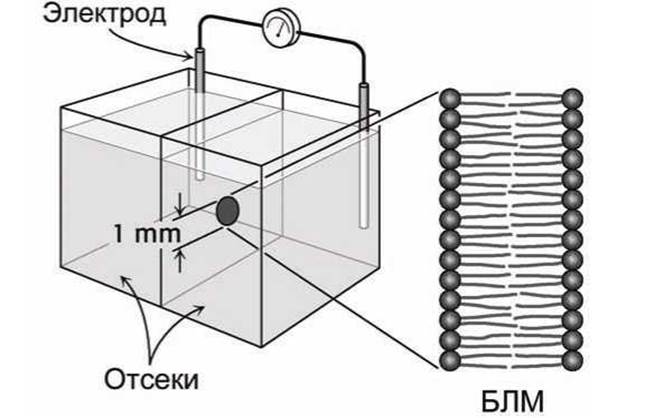
Figure 31 - Investigation of a planar Lipid Bilayer Membrane (BLM)
Liposome sizes typically range from a few tens of nanometers for small liposomes (consisting of a single bilayer, known as unilamellar vesicles, Figure 32) up to several hundred nanometers or even a micrometer for large multilamellar liposomes (consisting of many concentric bilayers).
The lipid bilayers of a multilamellar liposome are separated by aqueous compartments. Depending on The Nature of the Lipids, the thickness of the lipid layers is 6.5-7.5 nm, with an interlamellar distance of 1.5-2 nm. The overall diameter of multilamellar liposomes ranges from 60 nm to 400 nm or more.
Unilamellar liposomes can be prepared by various methods, such as sonicating a suspension of multilamellar liposomes. The diameter of unilamellar liposomes produced by this method is about 25-30 nm. Other preparation techniques have also been developed, yielding vesicles with diameters up to 400 nm and greater.
Liposomes serve as valuable models for investigating various properties of cellular biomembranes because they act, in a sense, as a prototype of a cell. They allow researchers to study numerous characteristics of natural membranes that are primarily related to the composition and state of the phospholipid phase. Biological membranes are composed of the same phospholipid bilayers, although they also incorporate numerous protein molecules.

Figure 32 - Unilamellar liposome
Liposomes are widely used in medicine. A therapeutic drug is encapsulated within the liposome, utilizing it as a phospholipid microcapsule to deliver the medication to specific Organs and Tissues. Liposomes are non-toxic (given the proper Selection of lipids), fully biodegradable by the body, and capable of crossing certain biological barriers.
For instance, Insulin encapsulated in a liposome is protected from the action of digestive Enzymes. Currently, researchers are exploring the possibility of administering liposomal drugs orally, which could free diabetes patients from The Need for regular injections.
Recent years have seen ongoing research into developing liposomal therapies for tumors, enzymatic deficiencies, and atherosclerosis. Scientists are studying the feasibility of targeted drug delivery, wherein liposome-encapsulated medications are directed to a diseased organ or even a specific pathological site (such as a damaged region of The Heart). To achieve this, an antibody protein specific to a membrane antigen of the target organ is attached to the liposome surface. The liposomes circulate throughout the body via the bloodstream and accumulate specifically when they reach the target organ.
METABOLISM/35.html">Review Questions and Exercises
1. What state of condensed matter is referred to as liquid-crystalline? List the various possible liquid-crystalline structures.
2. How does the presence and number of unsaturated C=C bonds in the hydrocarbon tails of lipids affect their melting point?
3. Characterize the phase transition of a biomembrane from the liquid-crystalline to the gel state.
4. What adaptive changes occur in The chemical composition of biomembranes during prolonged exposure to low ambient temperatures?
5. What is the primary mechanism underlying cryoinjury (Damage caused by cooling) in biological membranes?
6. How can the phase transition of a biomembrane from the liquid-crystalline to the gel state facilitate cellular thermoreception?
7. How does the presence of Cholesterol molecules affect the fluidity (viscosity) of a biomembrane?
8. Under what conditions does The addition of cholesterol molecules increase membrane thickness, and when does it not?
9. How does Lipid Composition influence the curvature of a biomembrane?
10. What modifications of lipid molecules are catalyzed by phospholipases?
11. What is the Functional Significance of the asymmetric distribution of phospholipids between the cytosolic and exoplasmic leaflets of a biomembrane?
12. For what purposes are model lipid membranes used?
13. What Three types of model membranes have found the most widespread application?
14. What are monomolecular lipid layers used for?
15. Describe the Langmuir-Blodgett method for producing multilayer oriented membranes.
16. Describe the Mueller method for producing planar bilayer lipid membranes.
17. What are planar bilayer lipid membranes used for?
18. What is the difference between unilamellar and multilamellar liposomes?
19. What are liposomes used for?
Last update: 13/08/2026
Editorial and Educational Adaptation: This material has been compiled based on the primary/original source text. The project team performed an editorial review, corrected technical inaccuracies, structured sections, and adapted the content for an educational format.
What was processed:
- elimination of formatting defects (OCR errors, structural breaks, corrupted characters);
- editorial organization of content;
- standardization of terminology in accordance with academic sources;
- verification of factual statements against the original source text.
All mentions of the author, publication year, and origin of the primary text have been preserved in accordance with the source.