Review of Medical Physiology - William F. Ganong 2002
Functions of the Nervous System
Regulation of Posture and Movement
Posture Control System - The Role of the Medulla Oblongata
In experimental animals where the Hindbrain and Spinal Cord are disconnected from the rest of the CNS by transecting the Brainstem at the upper border of the Pons, the primary manifestation of the lesion is marked spasticity of the body musculature. This surgical Procedure is called decerebration, and the characteristic spasticity that ensues is known as decerebrate rigidity. Decerebrate rigidity differs in its temporal course from spinal Shock, as the rigidity develops immediately upon transection of the brainstem.
Mechanism of Decerebrate Rigidity
The manifestation of decerebrate rigidity is spasticity resulting from the diffuse enhancement of stretch Reflexes. This phenomenon is driven by two factors: an increase in the overall excitability of the motoneuron pool and a corresponding shift in potentials within γ-efferent Neurons.
Supraspinal Regulation of Stretch Reflexes
The brainstem regions that send impulses either facilitating or inhibiting stretch reflexes are illustrated in Fig. 12-6. For the most part, these impulses act by either increasing or decreasing the sensitivity of Muscle spindles (Fig. 12-7). A sizable stimulatory area within the reticular Formation of the brainstem fires spontaneously, presumably driven by afferent impulses in a manner similar to the ARAS. However, a smaller brainstem area that inhibits excitation in γ-efferent neurons does not fire spontaneously; instead, it is driven by impulses descending from the Cerebral Cortex AND Cerebellum. The inhibitory area in the basal nuclei either acts directly via descending pathways, as shown in Fig. 12-6, or stimulates the cortical inhibitory center. Impulses from both the inhibitory and stimulatory areas of the reticular formation travel downward within the lateral funiculus of the spinal cord. When the brainstem is transected at the upper border of the pons, two of the three inhibitory areas controlling the reticular inhibitory center remain above the level of transection, and their influence is consequently abolished. The Influence of the stimulatory area remains intact; thus, the balance between stimulatory and inhibitory inputs acting on the γ-efferent neurons shifts toward excitation. The Membrane Potential in γ-efferent neurons increases and, As a result, stretch reflexes become hyperactive. Extirpation of the cerebellum in decerebrate animals—thereby removing yet another inhibitory region—further increases the degree of rigidity. The mechanisms of cerebellar influence are complex; for instance, cerebellar destruction in humans typically results in hypotonia rather than spasticity.
Class="center">
Fig. 12-6. Brain regions in the cat whose stimulation exerts a facilitating (plus sign) or inhibiting (minus sign) effect on stretch reflexes: 1, motor cortex; 2, basal nuclei; 3, cerebellum; 4, inhibitory area of the reticular formation; 5, stimulatory area of the reticular formation; 6, vestibular nuclei (reproduced with permission from Lindsley DB, Schreiner LH, Magoun HW: An electromyographic study of spasticity. J Neurophysiol 1949;12:197).
The vestibulospinal tract and certain other descending pathways also enhance stretch reflexes and contribute to rigidity. Unlike the pathways originating in the reticular formation, these descend primarily within the anterior funiculus of the spinal cord, and the rigidity resulting from their excitation cannot be abolished by muscle deafferentation. This indicates that such rigidity is caused by a direct excitatory action on γ-motoneurons that elevates their excitability, rather than by action mediated through the gamma motor system, which would otherwise be blockable by deafferentation.

Fig. 12-7. Responses of isolated afferent fibers from muscle spindles to varying degrees of muscle stretch. Numbers on the left indicate the degree of stretch. Upward deflections represent action potentials, and downward deflections represent stimulus artifacts. Recordings were made before brain stimulation (first Column), during stimulation of brain areas that facilitate (second column) or inhibit (third column) stretch reflexes, and after transection of motor nerves (fourth column) (reproduced with permission from Eldred E, Granit R, Merton PA: Supraspinal control of muscle spindles. J Physiol [Lond] 1953;122:498).
Nature of Decerebrate Rigidity
In dogs and cats, the spasticity resulting from decerebration predominantly affects extensor Muscles. Sherrington noted that these muscles counteract gravity in the animal; when placed in an upright position, the posture of a decerebrate cat or dog serves as a "caricature of normal standing."
Decerebration experiments have also helped to elucidate the mechanisms of tonic static reflexes that help support the animal's body against gravity. Further support for this interpretation comes from observations of arboreal animals. These creatures spend most of their time hanging upside down from branches and exhibit flexor muscle rigidity. In humans, as in experimental cats and dogs, true decerebrate rigidity is accompanied by increased extensor tone. Apparently, humans have not evolved far enough from their quadrupedal ancestors to develop fundamentally different response patterns in the upper extremities, even though the primary antigravity muscles in the standing posture are flexors. However, true decerebrate rigidity is rare in humans, and the lesions that cause it are usually fatal.
More commonly, rigidity of the lower extremity extensors and moderate flexion of the upper extremities appear as signs of decorticate rigidity, caused by damage to the cerebral cortex with an intact brainstem (Fig. 12-8).
Tonic Labyrinthine Reflexes
In decerebrate animals, the pattern of limb rigidity depends on the position in which the limbs are placed. Righting reflexes are absent, and the animals remain in whatever posture they are placed. If the animal is placed on its back, maximal extension is observed in all four limbs; when placed on either side, the rigidity decreases; and when placed prone, the rigidity is minimal. These changes in rigidity—known as tonic labyrinthine reflexes—are driven by the EFFECT OF GRAVITY on the otolith Organs and are mediated via vestibulospinal pathways. This effect is somewhat unexpected in terms of understanding the functional role of rigidity in standing, and its precise physiological significance remains unclear.
Compelling evidence for Structure/19.html">The Importance of the vestibular apparatus in postural regulation comes from the effects of labyrinthectomy in cats. Normally, a cat easily learns to traverse a rotating beam. Following unilateral labyrinthectomy, this ability is lost, but it recovers within about six weeks due to adaptive Changes in the remaining postural pathways. However, after bilateral labyrinthectomy, The ability to walk on a rotating beam is permanently lost.
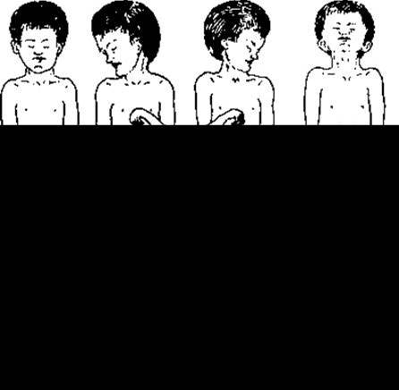
Fig. 12-8. Decorticate rigidity (A–C) and true decerebrate rigidity (D). A, Patient lying supine with HEAD unrotated; B and C, tonic neck reflex elicited by turning the head to the right or left (reproduced with permission from Fulton JF [editor]: Textbook of Physiology, 17th ed. Saunders, 1955).
Tonic Neck Reflexes
Changing THE POSITION OF the head relative to the trunk in decerebrate animals alters the pattern of rigidity. When the head is turned to one side, the limbs on that side (the "jaw limbs") extend and become more rigid, whereas the rigidity of the contralateral limbs decreases. This posture is also characteristic of intact animals turning the head to look sideways. Flexion of the head causes flexion of the forelimbs and extension of the hindlimbs, producing a posture resembling that of an animal peering into a burrow. Extension of the head causes flexion of the hindlimbs and extension of the forelimbs, producing a posture typical of an animal looking over an obstacle. These reactions are called tonic neck reflexes. They are initiated by proprioceptors responding to muscle stretch in the upper neck region and can persist for prolonged periods.
Last update: 10/08/2026
Editorial and Educational Adaptation: This material has been compiled based on the primary/original source text. The project team performed an editorial review, corrected technical inaccuracies, structured sections, and adapted the content for an educational format.
What was processed:
- elimination of formatting defects (OCR errors, structural breaks, corrupted characters);
- editorial organization of content;
- standardization of terminology in accordance with academic sources;
- verification of factual statements against the original source text.
All mentions of the author, publication year, and origin of the primary text have been preserved in accordance with the source.