Review of Medical Physiology - William F. Ganong 2002
Respiration
Pulmonary Functions
Anatomy of the Respiratory System
Airways
Inhaled air is warmed and humidified as it passes through the Nasal cavity and Pharynx. It then travels through the Trachea, Bronchi, bronchioles, respiratory bronchioles, alveolar ducts, and finally reaches the alveoli (Fig. 34-1). Between the trachea and the alveoli, the airways divide 23 times. The first 16 generations form the conducting zone of the airways, which merely transports gas to and from the external environment. This zone comprises the bronchi, bronchioles, and terminal bronchioles. The remaining seven generations constitute the transitional and respiratory zones proper, where gas exchange takes place. These include the respiratory bronchioles, alveolar ducts, and alveoli. This multiple structural branching dramatically increases the total cross-sectional area of the airways: from 2.5 cm2 in the trachea to 11,800 cm2 in the alveoli (Fig. 34-2). This is why the velocity of airflow decreases only slightly, even in small-diameter airways.
Class="center">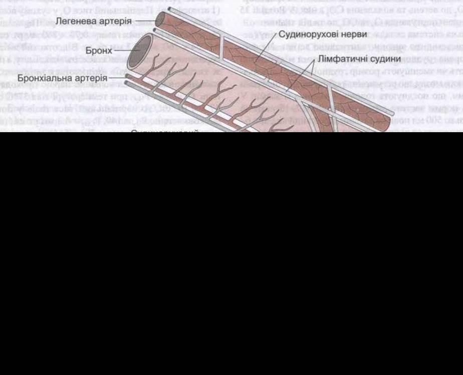
Fig. 34-1. Structure OF THE lung: A - alveolus; AP - alveolar duct; RB - respiratory bronchiolar; TB - terminal bronchiole (modified from Staub NC: The Pathophysiology of pulmonary edema. Hum Pathol 1970;1:419).
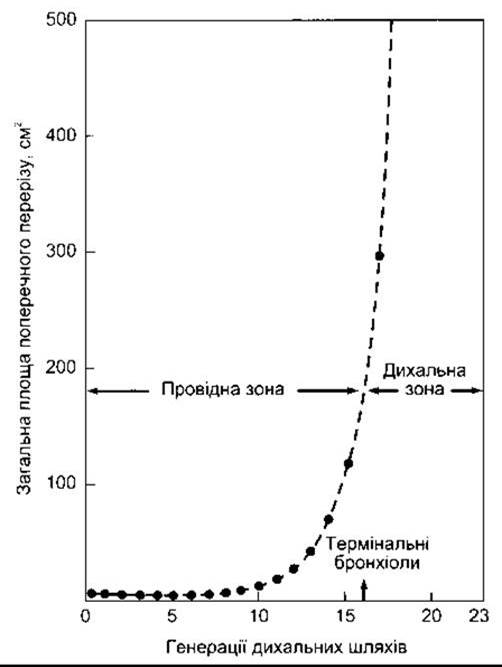
Fig. 34-2. Relationship between total cross-sectional area and airway generations. Note how rapidly the total cross-sectional area increases in the respiratory zone. Consequently, the linear velocity of gas during inhalation drops to a very low level in this zone (reproduced with permission from West JB: Respiratory Physiology: The Essentials, 4th ed. Williams & Wilkins, 1990).
The alveoli are surrounded by pulmonary capillaries. Consequently, air and Blood are separated only by the alveolar epithelium and capillary endothelium, with a combined thickness of approximately 0.5 µm (Fig. 34-3). The Human Body contains 300 million alveoli, and the total surface area of their walls in contact with capillaries exceeds 70 m2 in both Lungs combined.
The alveoli consist of Two Types of epithelial Cells. Type I alveolar cells (type I pneumocytes) are squamous epithelial cells with extensive, extended Cytoplasm closely apposed to the capillaries. Type II alveolar cells (granular pneumocytes) are thinner and contain characteristic cytoplasmic inclusion bodies. These cells produce surfactant (see below). Other Cell types can also be identified in the alveoli, typically alveolar macrophages (AMs), lymphocytes, plasma cells, APUD cells (see Chapter 26), and mast cells.
Mast cells (see Chapter 27) contain heparin, Lipids, histamine, and proteases, which are involved in allergic reactions.
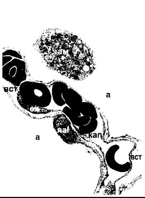
Fig. 34-3. Region of the interalveolar septum in an adult human lung. A pulmonary capillary (cap) within the septum contains plasma and erythrocytes. Note the closely apposed endothelial wall and pulmonary epithelium, separated in places by Connective Tissue fibers (ctf); en - Nucleus of the endothelial cell; tpI - nucleus of the type I alveolar cell; a - alveolar space; am - alveolar macrophage (reproduced with permission from Burri PA: Development and growth of human lung. In: Handbook of Physiology, Section 3, The Respiratory system. Fishman AP, Fisher AB [editors]. American Physiological Society, 1985).
Bronchi and Their Innervation
The walls of the Trachea and Bronchi consist of Cartilage and a relatively small amount of smooth Muscle. Their inner surface is lined with ciliated epithelium and contains mucous and serous glands. The cilia extend all the way down to the respiratory bronchioles. However, neither glands nor cartilage are found within the epithelium of the bronchioles and terminal bronchioles. In contrast, these regions contain a significantly higher proportion of smooth muscle, especially considering the muscle mass involved in thinning the bronchiolar walls. The walls of the bronchi and bronchioles are innervated by the Autonomic Nervous system. A high density of muscarinic receptors is present here, and activation of these cholinergic receptors induces bronchoconstriction. β2-Adrenergic receptors are located in the bronchial epithelium and smooth muscle. A certain portion of bronchial smooth muscle lacks direct innervation. Some of these fibers, located near cholinergic nerve terminals, neutralize acetylcholine. Stimulation of β2-adrenergic receptors causes bronchodilation along with an increase in bronchial secretion (see Table 13-2), whereas α1-adrenergic receptors inhibit this process. Furthermore, there is an additional non-cholinergic, non-adrenergic (NANC) innervation in the bronchioles that also promotes bronchodilation; there is also evidence that VIP acts as a bronchodilatory neurotransmitter.
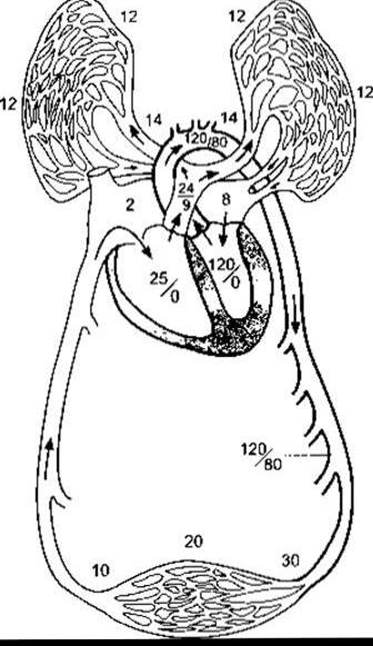
Fig. 34-4. Blood pressure (mmHg) in the pulmonary and systemic circulations (modified and reproduced with permission from Comroe JH Jr: Physiology of Respiration, 2nd ed. Year Book Medical Publishers, 1974).
Virtually all of the body's blood passes through the pulmonary Arteries and pulmonary capillary bed, where it is oxygenated, and then returns via the Pulmonary Veins to the left atrium (Fig. 34-4). Specialized, much smaller-diameter bronchial arteries branch off from the systemic arteries and form capillaries that either drain into the bronchial veins or anastomose with pulmonary capillaries or pulmonary veins (Fig. 34-5). The bronchial veins empty into the azygos vein. The bronchial circulation supplies nutrients to the bronchi and Pleura. Lymphatic vessel density here is among the highest of any organ (see Fig. 34-1).
Other Aspects of the pulmonary circulation are described below.
Last update: 10/08/2026
Editorial and Educational Adaptation: This material has been compiled based on the primary/original source text. The project team performed an editorial review, corrected technical inaccuracies, structured sections, and adapted the content for an educational format.
What was processed:
- elimination of formatting defects (OCR errors, structural breaks, corrupted characters);
- editorial organization of content;
- standardization of terminology in accordance with academic sources;
- verification of factual statements against the original source text.
All mentions of the author, publication year, and origin of the primary text have been preserved in accordance with the source.