Medical Radiology - Lazar A.P. 2008
Radiation examination of the heart and blood vessels
To study the morphological and functional state of the circulatory Organs, the main Diagnostic imaging Methods are used: X-ray imaging, ultrasound, computed tomography, Magnetic Resonance Imaging, and radionuclide scanning.
X-ray examination includes fluoroscopy, radiography, and contrast injection methods such as angiocardiography, ventriculography, aortography, coronarography, angiography, and digital subtraction angiography.
Fluoroscopy allows the evaluation of the position, shape, and size of The Heart, as well as the pulsation of its chambers and great vessels. Pulsation is most pronounced along the contours of the ventricular arches.
To reduce projection magnification and preserve the sharpness of the pulmonary pattern, radiography is performed at a focal distance of 1.5–2 m (teleradiography) with a minimal exposure time (0.1 s). Radiography and fluoroscopy of the heart are performed in four standard projections: anteroposterior (straight), right (first) oblique, left (second) oblique, and left lateral. In the direct anteroposterior projection, the patient stands facing the screen (or the film cassette), pressing firmly against it with the anterior chest wall, with hands resting on the hips and elbows brought forward as much as possible to pull the scapulae out and prevent them from obscuring the lung fields; the patient's shoulders are lowered. In the right oblique position, the patient stands with the right shoulder forward at an angle of approximately 450, hands remain on the hips, the left elbow is brought forward as much as possible while the right is pulled back, and shoulders are lowered. Similarly to the right oblique position, in the left oblique position the patient stands at an angle of 450 with the left shoulder forward. In the left lateral projection, the subject stands with their left side facing the screen or X-ray film so that the shadow of the Sternum anteriorly forms the outer contour, with hands positioned behind the HEAD.
Radiological examination begins with studying the shape and condition of the thoracic Skeleton, the position and shape of the diaphragmatic domes, the transparency of the lung fields, the pattern of the pulmonary vasculature, and the lung roots. After that, the examination proceeds to the heart itself and the great vessels.
To examine the shape and size of the left atrium, a contrast Study of the Esophagus in the right oblique projection is often used, during which the patient swallows one teaspoon of a barium suspension, and radiographs are taken as the contrast agent passes into The Stomach. The contrast medium remaining between the folds of the esophageal mucosa outlines the posterior surface of the left atrium as a narrow shadow strip.
Contrast injection methods for imaging The Heart and great vessels are performed in specialized cardiosurgical clinics. These methods are used to diagnose intracardiac Blood flow disorders in congenital and acquired defects of the heart and great vessels. High-density iodinated Water-soluble agents are used for contrast enhancement: ionic (urographin, cardiotrast, verographin, triombrast) and non-ionic (omnipaque, ultravist).
Class="center">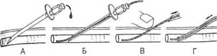
Fig. 134. Performance of Seldinger arteriography.
A — arterial puncture with a trocar;
B — insertion of a metallic guidewire through the trocar;
C — withdrawal of the trocar;
D — advancement of the catheter over the metallic guidewire.
During general angiocardiography, a radiopaque contrast agent is injected into the median cubital vein using an automatic injector at a rate of 45 ml/s based on 1 ml of solution per 1 kg of the patient's body weight. Serial angiocardiograms taken with specialized high-speed equipment (10–12 frames per second) or via cineangiocardiography (24–48 frames per second) provide sequential images of the basilic, brachial, axillary, subclavian, brachiocephalic, and SUPERIOR VENA CAVA Veins, followed by the right atrium, right ventricle, pulmonary trunk, pulmonary Arteries, and later the Pulmonary veins, left atrium, left ventricle, with the contrasted aorta visible on the final frames. In selective angiocardiography, the median cubital vein is punctured, a catheter is advanced under fluoroscopic guidance into the right atrium or ventricle, and the contrast agent is injected. To contrast the left side of the heart, puncture of the left atrium can be performed from the right atrium through the interatrial septum. Compared to general angiography, selective angiography provides sharper contrast of individual heart chambers and reduces the volume of administered radiopaque contrast medium.
For ventriculography or aortography, femoral artery puncture is performed using the Seldinger technique (Fig. 134), a catheter is advanced into the left ventricle or aorta accordingly, a radiopaque contrast agent is injected, and a series of radiographs is obtained. These methods are used to detect ventricular septal defects, mitral regurgitation, aortic valve regurgitation, congenital Anomalies of the aorta and its branches, aortic coarctation (narrowing), etc.
Coronarography is a contrast examination of the coronary arteries used to detect the localization, extent, and degree of arterial stenosis, as well as to evaluate the state of collateral Circulation. Coronarography is performed via percutaneous puncture of the femoral artery, advancement of a catheter to the aortic sinuses, and selective catheterization of the right or left coronary arteries. During coronary catheterization, it is possible to perform intravascular dilation, which is the widening of narrowed segments of the coronary arteries.
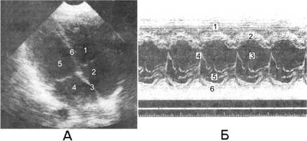
Fig. 135. The heart during Ultrasound examination.
A — two-dimensional echocardiography: 1 — right ventricle; 2 — right atrium; 3 — interatrial septum; 4 — left atrium; 5 — left ventricle; 6 — interventricular septum;
B — M-mode echocardiography: 1 — right ventricle; 2 — interventricular septum; 3 — left ventricle; 4 — anterior mitral leaflet; 5 — posterior mitral leaflet; 6 — posterior wall of the left ventricle.
Angiography is used to study the condition of Blood Vessels: arteries (arteriography), veins (venography or phlebography), and Lymphatic vessels (lymphography). Digital subtraction angiography involves acquiring images using a computer that subtracts the pre-contrast image from the image obtained after the injection of contrast into the vessel. This eliminates the Background shadow of surrounding organs, improving image quality of the contrasted vessels and reducing the required amount of contrast medium.
Ultrasound examination of the heart (echocardiography) is an informative, inexpensive, and safe real-time diagnostic method, which is why it has become the primary modality for cardiac imaging. M-mode echocardiography allows obtaining a one-dimensional display of the motion of individual cardiac structures throughout the cardiac cycle. The echogram appears as a set of curves, each corresponding to a specific heart Structure: the Pericardium, ventricular and atrial walls, interatrial and interventricular septa, and Valves (Fig. 135). Two-dimensional (2D) echocardiography (B-mode) enables the evaluation of the anatomy, function, and pathological conditions of the heart chambers and valves, the interventricular septum, myocardial contractility, and pericardial pathology. Doppler echocardiography studies the direction and velocity of blood flows, making it possible to detect intracardiac shunts and valvular lesions. Duplex Ultrasonography, which combines Doppler sonography with two-dimensional imaging (B-mode), evaluates vascular status alongside blood flow analysis in any segment of a vessel.

Fig. 136. X-ray Computed Tomography scan of the thoracic cavity in the horizontal plane with intravenous contrast enhancement.
1 — left ventricle;
2 - left atrium;
3 - right atrium;
4 - mitral valve;
5 - right ventricle;
6 - interventricular septum.
Computed tomography allows obtaining cross-sectional images of the heart with a thickness of 2 mm or less in a relatively short time. To enhance the diagnostic capabilities of computed tomography, contrast enhancement techniques involving intravenous administration of radiopaque agents are used (Fig. 136). CT is used to determine the dimensions of individual heart chambers, the condition of major vessels and coronary vessels, and the presence of fluid in the pericardial cavity. This enables the detection of cardiac tumors, aortic aneurysms, coronary artery stenosis and atherosclerosis, thrombi, pericarditis, and Congenital heart defects. CT often employs an invasive contrast enhancement technique following intravenous bolus injection of water-soluble iodine-containing agents. Advanced CT modalities, such as ultrafast CT or cine-CT, are effective for the quantitative assessment of ventricular dimensions and function, cardiac hemodynamics, patency of aortocoronary bypass grafts, and complications of myocardial infarction. Modern high-speed helical computed scanners and specialized software enable the generation of three-dimensional (3D) models of the heart during various Phases of the cardiac cycle.
Magnetic resonance imaging is typically performed using spin-echo pulse sequences synchronized with ECG gating, which permits high-resolution evaluation of cardiac Morphology. Recently, cine-MRI has been widely adopted, allowing a single MR image to be acquired within one second. This facilitates the visualization of contrast agent dynamics within the heart chambers, enabling the assessment of blood volume, velocity, and flow direction, myocardial contractility, myocardial perfusion, and valvular function.
Radionuclide imaging most commonly involves the intravenous administration of 99mTc-pertechnetate. The myocardial uptake of this radiopharmaceutical peaks within 5–10 minutes. This method provides valuable data on ventricular function, myocardial perfusion, and the presence of myocardial infarction (visualized as areas of reduced radionuclide uptake).

Fig. 137. Cardiac contours in various projections.
A - horizontal cross-section; B - left lateral projection; C - right oblique projection; D - anterior anteroposterior projection; E - left oblique projection.
Ao - aorta;
SVC - superior vena cava;
LAA - left atrial appendage;
PT - pulmonary trunk;
LA - left atrium;
LV - left ventricle;
RA - right atrium;
RV - right ventricle; Esoph - esophagus.
Last update: 08/08/2026
Editorial and Educational Adaptation: This material has been compiled based on the primary/original source text. The project team performed an editorial review, corrected technical inaccuracies, structured sections, and adapted the content for an educational format.
What was processed:
- elimination of formatting defects (OCR errors, structural breaks, corrupted characters);
- editorial organization of content;
- standardization of terminology in accordance with academic sources;
- verification of factual statements against the original source text.
All mentions of the author, publication year, and origin of the primary text have been preserved in accordance with the source.