Medical Radiology - Lazar A.P. 2008
Imaging Examination of the Chest Organs
Radiation Anatomy of the Chest Organs
During an X-ray Examination of the chest Organs (clinically abbreviated as CXR), the state of the Skeleton, the soft Tissues of the chest wall, and the condition of the thoracic cavity organs—the Lungs and Mediastinum—are evaluated. On a standard anteroposterior (AP) chest radiograph, the upper 7 pairs of Ribs are visible along almost their entire length. The lower ribs are partially obscured by the shadow of the Diaphragm and abdominal organs. The posterior PARTS OF THE ribs are narrower, oriented more horizontally, and cast a more intense shadow due to their greater thickness. The cartilaginous parts of the ribs do not cast a noticeable shadow on radiographs. Only after the age of 30, starting with the 1st costal Cartilage, does calcium deposit within them in the form of narrow bands along the margin and small islands in the center of the cartilage.
The scapula and clavicle are clearly visible under fluoroscopy and on the radiograph (Fig. 96). When the patient is correctly positioned in the AP projection, the sternal ends of the clavicles are located symmetrically at an equal distance from the spinal Column. The shadow of the Sternum in the AP projection is invisible because it merges with the shadow of the mediastinum and thoracic vertebrae; in the lateral projection, the sternum forms the anterior contour. On a high-quality radiograph, the upper 3–4 thoracic vertebrae can be distinguished, while the lower vertebrae are obscured by the mediastinal shadow.
On both sides of the chest, the edge of the pectoralis major Muscle is visible, which is more pronounced in men. In women, the shadows of the Mammary Glands are superimposed on the lower lung fields, with the lower edge of the breast appearing as an arcuate shadow. In both women and men, symmetrical shadows of the nipples are often visible, which can be mistakenly interpreted as pathological opacities.
The diaphragm, together with the abdominal organs, bounds the clear lung fields from below as a distinct, arcuate, dense shadow. Only to the left of the midline, beneath the contour of the diaphragm, can the radiolucency of the gastric air bubble be seen. The right dome of the diaphragm is located approximately at the level of the anterior end of the 6th rib, and the left is one rib lower.
Class="center">
Fig. 97. Radiographs of the chest organs in the anteroposterior (A) and left lateral (B) projections.
In the lateral angles of the diaphragmatic domes, the costodiaphragmatic pleural recesses (or sinuses) are visible, and in the medial angles, the phrenicomediastinal recesses (Fig. 97). In the lateral projection, the anterior costodiaphragmatic recesses are located higher than the posterior ones.
In the AP projection, the following lung fields are distinguished within each lung: 1) apical, located above the clavicles; 2) upper, from the clavicle to the anterior section of the 2nd rib; 3) middle, from the 2nd to the 4th rib; 4) lower, from the 5th rib to the diaphragm.
In the lateral projection, the shadows of the sternum, scapulae, and 3rd–9th thoracic vertebrae are clearly visible, from which the shadows of the ribs extend obliquely downward. The shadows of the ribs closest to the cassette are narrower and sharper. The translucent areas between the sternum and The Heart with the aorta are called the retrosternal space, and those between The Heart and the spinal column are called the retrocardiac space.

Fig. 98. Structural components of the lung roots. A — Anatomical Structure; B — diagram of the radiographic pattern.
1 — left main bronchus;
2 — pulmonary trunk;
3 — left atrium;
4 — right pulmonary artery.

Fig. 99. Projection of lobes and interlobar fissures on the radiograph (diagram).
A — right lateral projection; B — anteroposterior projection; C — left lateral projection.
1 — upper lobe; 2 — middle lobe; 3 — lower lobe; 4 — horizontal fissure; 5 — oblique fissure.
The shadow of the lung ROOT is formed by large pulmonary Arteries, Veins, Bronchi, and to a lesser extent, Lymphatic vessels and nodes (Fig. 98). In the anterior projection, this shadow is heterogeneous (structured), located at the level of the anterior ends of the 2nd–4th ribs, about 2 cm wide, and occupies no more than a quarter of the transverse dimension of the lung. The root of the left lung is located higher than the right, and its lower part is obscured by the cardiac shadow. In the lateral projection, the lung roots lie beneath the aortic arch.
The largest Lymph Nodes of the thoracic cavity are located in the lung roots and mediastinum. In inflammatory and tumor processes, they enlarge, and the shadow of the root becomes unstructured.
The pulmonary pattern refers to the linear shadows of the pulmonary vessels extending from the lung roots, which branch and intertwine to form a reticular structure. The caliber of these shadows gradually decreases toward the periphery, stopping about 2 cm short of the chest wall.
The parietal Pleura can sometimes be traced in the AP projection as a thin arcuate strip extending from the apex along the lateral surface of the lung. The interlobar pleura and the clear boundary between the lung lobes are not discernible in the AP projection. The AP PROJECTION OF THE oblique fissure runs from the 3rd thoracic vertebra obliquely lateral and downward to the lateral surface of the 4th rib, and then obliquely downward and medially to the diaphragm. The horizontal fissure of the right lung runs horizontally inward from the lateral surface of the 4th rib (Fig. 99). In the lateral projection, the pleura of the oblique interlobar fissure forms a thin "hairline" shadow extending obliquely downward and forward from the level of the 4th thoracic vertebra to the diaphragm. In the lateral projection, the horizontal fissure starts from the oblique fissure at the level of the lung root and runs almost horizontally forward to the chest wall.

Fig. 100. Branching of segmental bronchi:
A – right lateral projection; B – posteroanterior projection; C – left lateral projection.
1 – apical; 2 – posterior; 3 – anterior; 4 – lateral (upper lingular in the left lung); 5 – medial (lower lingular in the left lung); 6 – superior; 7 – medial basal (often absent in the left lung); 8 – anterior basal; 9 – lateral basal; 10 – posterior basal; 11 – horizontal fissure; 12 – oblique fissure.
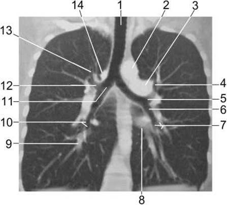
Fig. 101. Contrast-enhanced computed tomography scan of the chest; coronal reconstruction.
1 – Trachea; 2 – aortic arch; 3 – left pulmonary artery; 4 – apicoposterior segmental bronchus; 5 – left upper lobar bronchus; 6 – left lower lobar bronchus; 7 – left basal segmental bronchi; 8 – left inferior pulmonary vein; 9 – right inferior pulmonary vein; 10 – right basal segmental bronchi; 11 – right main bronchus; 12 – right upper lobar bronchus; 13 – apical segmental bronchus; 14 – azygos arch.
The lung lobes consist of segments shaped as irregular pyramids, with their apices directed toward the roots and their bases toward the pleural surface. While the boundaries between bronchopulmonary segments are not visible on conventional radiographs, understanding the Topographic anatomy of the bronchial tree, lobes, and segments is essential for localizing pathological processes. The right lung comprises 3 lobes and 10 segments, whereas the left lung consists of 2 lobes and 10 (or 9) segments (Figs. 100, 101).
The spatial relationships of the superior mediastinal organs on axial computed tomography slices of the thoracic cavity are demonstrated in Figures 102 and 103.
Last update: 08/08/2026
Editorial and Educational Adaptation: This material has been compiled based on the primary/original source text. The project team performed an editorial review, corrected technical inaccuracies, structured sections, and adapted the content for an educational format.
What was processed:
- elimination of formatting defects (OCR errors, structural breaks, corrupted characters);
- editorial organization of content;
- standardization of terminology in accordance with academic sources;
- verification of factual statements against the original source text.
All mentions of the author, publication year, and origin of the primary text have been preserved in accordance with the source.