Medical Radiology - Lazar A.P. 2008
Radiation examination of bones and joints
Joint diseases
Based on their Etiology, Joint Diseases are classified into infectious (Arthritis) and degenerative-dystrophic disorders (arthrosis).
Infectious joint diseases are most commonly a complication of systemic infections, such as rheumatism, Influenza, measles, diphtheria, scarlet fever, brucellosis, Gonorrhea, etc.
Class="center">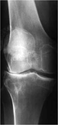
Fig. 54. Radiograph showing arthritis of the knee joint. Note the uneven narrowing of the joint space and subluxation.
Such cases are referred to as primary arthritis, where the infection spreads via a hematogenous route. In pediatric infectious diseases, infectious arthritis typically manifests during the convalescence period, most frequently affecting the hip joint. Secondary arthritis develops when the pathological process extends to the joint by direct contact.
Depending on the number of affected joints, arthritis is categorized as monoarthritis or polyarthritis. Based on The Nature of the effusion, it is divided into purulent (serous-purulent, purulent effusion) and non-purulent (serous, serous-fibrinous, serous-hemorrhagic effusion) forms.
By its pathway of spread, Purulent Arthritis can be primary, originating from a distant focus of infection, or secondary, resulting from adjacent Osteomyelitis. It is caused by the same pathogens responsible for osteomyelitis. The disease onset is acute, accompanied by high fever and systemic toxicity. Within 1-2 days, local symptoms emerge, including severe pain, Swelling, hyperemia, and localized hyperthermia of the joint. Purulent arthritis progresses through three phases: synovitis, capsular Phlegmon, and panarthritis. In the synovitis phase, radiographic findings mirror those of non-purulent arthritis, showing soft tissue swelling, obscuration of the physiological joint translucencies, widening of the joint space, and subluxation. In the capsular phlegmon phase, alongside the signs of synovitis, fistulous tracts discharging pus may form. Radiographically, soft tissue swelling is observed, while the height of the joint space progressively decreases (Fig. 54) due to articular Cartilage destruction and purulent discharge drainage. Marginal erosions measuring 0.5–1.0 cm with indistinct, irregular contours appear at the sites of Joint Capsule attachment to the bones As a result of purulent dissolution. Panarthritis is characterized by the extension of the pathological process from the capsule to the articular surfaces of the bones. The joint space is significantly narrowed, showing areas of osteolysis along the subchondral plate with compromised continuity and a serrated appearance (Fig. 55), accompanied by periostitis at the metaphyseal level. Further disease progression leads to osteomyelitis of the adjacent bone segments, whereas abatement of the process results in fibrous and, subsequently, bony ankylosis.
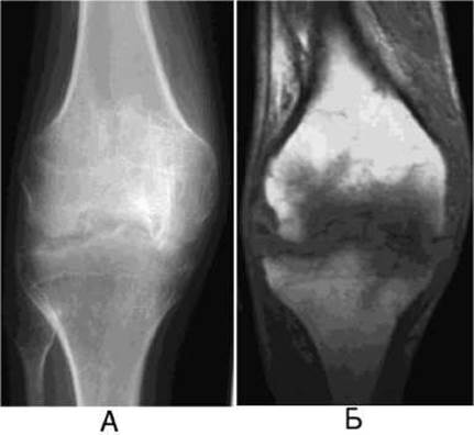
Fig. 55. Purulent arthritis of the knee joint.
A — radiograph demonstrates a reduction in joint space height, disrupted continuity and serration of the subchondral plate, and erosion of the articular bone surfaces;
B — magnetic Resonance image reveals a periarticular hypointense zone.
In rheumatic fever, joint involvement presents as an infectious-allergic inflammation. Acute rheumatic arthritis is more prevalent among young adults and children. Clinically, it is characterized by high BODY Temperature AND symmetrical pain and erythema in the large joints. Radiographic findings are often unremarkable or show mild Osteoporosis. Destructive Changes in the cartilage and articular bone surfaces are typically absent, and destruction foci in the epiphyses are rarely observed. Large joint effusions can occasionally lead to spontaneous, so-called distension subluxation or dislocation.
Rheumatoid arthritis (polyarthritis) ranks second in incidence after rheumatic fever. It predominantly develops in adults aged 30–50, with a female-to-male ratio of approximately 2:1. Its etiology remains unknown. Initially, the small JOINTS OF THE hands or feet are symmetrically affected, after which the process gradually extends to the large joints of the extremities. The onset of the disease is usually insidious in adults, but acute in children. Radiography reveals regional epiphyseal osteoporosis, initial widening of the joint spaces followed by subsequent narrowing, joint deformities, marginal bone defects, dislocations, subluxations, and ankylosis (Fig. 56). Subcutaneous nodules formed in rheumatoid arthritis do not undergo calcification and are not detectable radiographically.
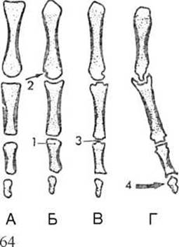
Fig. 56. Radiographic stages of rheumatoid arthritis (diagram).
A — normal radiograph;
B — early stage of the disease;
C — disease progression;
D — late Stages of the disease.
1 — band of radiolucency due to regional osteoporosis;
2 — marginal defects;
3 — decreased joint space height;
4 — joint destruction and dislocations.
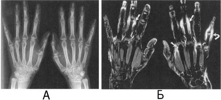
Fig. 57. Rheumatoid arthritis on a radiograph (A) and a magnetic resonance image (B).
In the Cytology/cytology/16.html">Early stages of the disease, when radiographic changes may be interpreted as minor deviations from the norm, osteoscintigraphy registers an increased radiopharmaceutical uptake in the joint area, while MRI reveals changes in the joints and surrounding soft Tissues (Fig. 57). Ultrasound of large joints makes it possible to determine the presence and amount of effusion in the early stages of the disease, as well as synovial thickening. MSCT allows for densitometric Assessment of the degree of osteoporosis.
The progression of rheumatoid arthritis leads to ulnar deviation of the fingers (Fig. 58) and their "swan-neck" deformity.
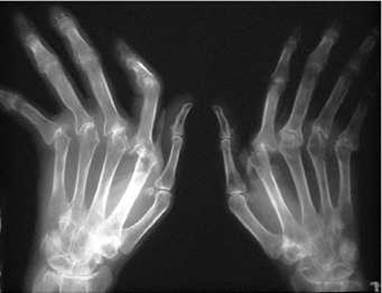
Fig. 58. Rheumatoid arthritis in the late stage of the disease on a radiograph.
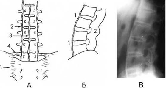
Fig. 59. Ankylosing spondylitis of the lumbar spine.
A - schematic diagram of an anteroposterior radiograph: 1 - sacroiliac ankylosis; 2 - numerous intervertebral ossifications ("railroad track" sign); 3 - calcification of the interspinous ligaments; 4 - calcification of the iliolumbar ligament.
B - schematic diagram of a lateral radiograph: 1 - intervertebral bone bridge; 2 - interapophyseal ankylosis.
C - lateral radiograph.
Gonococcal Arthritis is rare. Most commonly, a single joint is affected, more frequently the knee or wrist. Clinically, at the height of the disease, the joint exhibits acute pain, swelling, and elevated temperature. Radiologically, it presents with osteoporosis, irregular narrowing or complete disappearance of the joint space, uneven and hazy articular bone surfaces, and occasionally marginal erosions.
Ankylosing spondylitis, or Bechterew-Strümpell-Marie disease, typically manifests at the age of 20–25, predominantly in men. The condition develops gradually. Clinically, patients experience lower back pain; early on, a characteristic feature is a straight, rigid spine caused by the calcification of longitudinal ligaments, which gives the spine a "bamboo spine" appearance. The pathological process begins in the sacroiliac joint, subsequently spreading to the intervertebral and costovertebral junctions of the lumbar, thoracic, and cervical spine, and later involving the large joints, specifically the hip and knee. Chondritis and synovitis develop within the joints, progressing to fibrous and bony ankylosis. The Fibrous Connective Tissue eventually undergoes calcification, culminating in complete ankylosis.
Early radiological signs of ankylosing spondylitis are detected in the sacroiliac joint: progressive narrowing and obliteration of the joint space with The formation of bony ankylosis, and bone trabeculae bridging from the ilium to the sacrum. Later in the spinal Column, osteoporosis and the "railroad track" sign are observed—two parallel linear shadows running along both sides of the spine, caused by the calcification of the anterior and posterior longitudinal ligaments (Fig. 59).
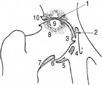
Fig. 60. Hip osteoarthritis on a radiograph (diagram).
1 - narrowing of the radiological joint space;
2, 4, 5 - inner and lower areas of osteosclerosis of the acetabulum;
3 - perifoveal osteophyte;
6 - pericapitis osteophyte;
7 - inferior osteophyte of the femoral neck;
8 - sclerosis of the upper part of the femoral HEAD and the acetabular roof;
9 - subchondral cyst;
10 - osteophyte of the acetabular roof.
Last update: 08/08/2026
Editorial and Educational Adaptation: This material has been compiled based on the primary/original source text. The project team performed an editorial review, corrected technical inaccuracies, structured sections, and adapted the content for an educational format.
What was processed:
- elimination of formatting defects (OCR errors, structural breaks, corrupted characters);
- editorial organization of content;
- standardization of terminology in accordance with academic sources;
- verification of factual statements against the original source text.
All mentions of the author, publication year, and origin of the primary text have been preserved in accordance with the source.