Human Anatomy - Kotsan I. Ya. 2009
Peripheral nervous system
Cranial nerves
Oculomotor nerve (III)
The Oculomotor nerve (n. oculomotorius) is a mixed nerve (Figs. 239, 255). It contains both motor and parasympathetic fibers. One portion of the nerve fibers originates from the motor Nucleus—the oculomotor nucleus (nucleus nervi oculomotorii), which is located beneath the ventral wall of the cerebral aqueduct at the level of the superior colliculi of the Midbrain tectum, while the other portion arises from the autonomic (parasympathetic) nucleus—the accessory oculomotor nucleus (nucleus oculomotorius accessorius), situated adjacent to the oculomotor nucleus. The nerve emerges from the Brain substance at the oculomotor sulcus of the cerebral peduncle and appears at the Base of the brain within the interpeduncular fossa. Extending anteriorly, the oculomotor nerve runs through the lateral wall of the cavernous sinus and then enters the orbital cavity via the superior orbital fissure.
Before entering the orbital cavity, the nerve divides into superior and inferior branches. The superior branch (ramus superior) is purely motor, innervating the levator palpebrae superioris Muscle and the superior rectus muscle of the eye. The inferior branch (ramus inferior) is mixed, supplying motor fibers to the inferior and medial rectus Muscles of the eye, as well as to the inferior oblique muscle. In addition, the oculomotor ROOT (radix oculomotoria) branches off from the inferior branch, consisting of axons from the parasympathetic nucleus Cells, and courses toward the ciliary ganglion. Autonomic fibers pass from the ciliary ganglion to innervate the smooth muscles of the eye: the sphincter pupillae and the ciliary muscle.
Class="center">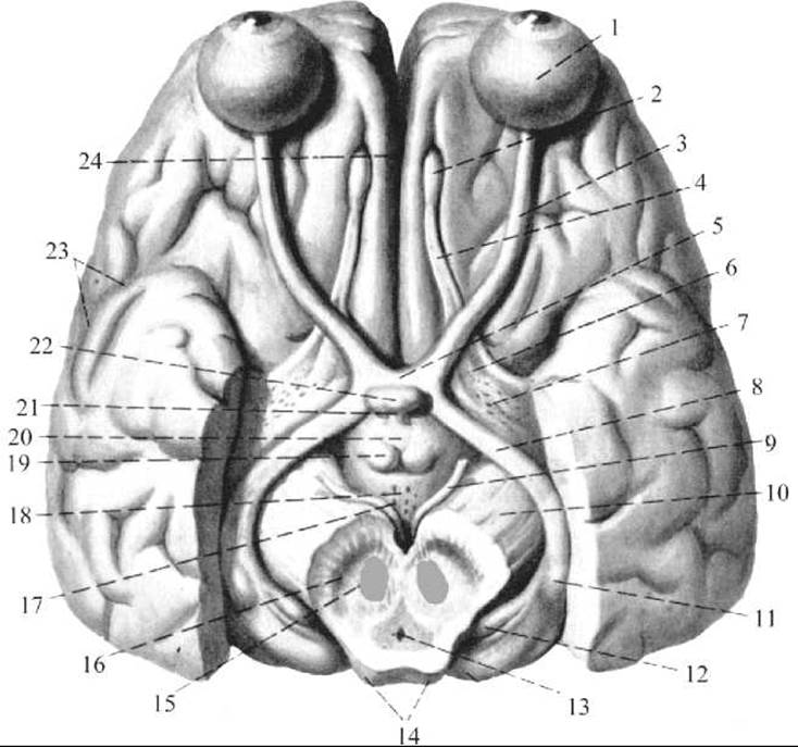
Fig. 254. Optic nerves and optic tracts (after R. D. Sinelnikov)
1 — Eyeball; 2 — olfactory bulb; 3 — Optic nerve; 4 — olfactory tract; 5 — optic chiasm; 6 — olfactory trigone; 7 — anterior perforated substance; 8 — optic tract; 9 — oculomotor nerve; 10 — cerebral peduncle; 11 — lateral geniculate body; 12 — medial geniculate body; 13 — cerebral aqueduct; 14 — midbrain tectum; 15 — red nucleus; 16 — substantia nigra; 17 — interpeduncular fossa; 18 — posterior perforated substance; 19 — mammillary body; 20 — tuber cinereum; 21 — infundibulum; 22 — Pituitary Gland; 23 — lateral sulcus; 24 — longitudinal cerebral fissure
Last update: 08/08/2026
Editorial and Educational Adaptation: This material has been compiled based on the primary/original source text. The project team performed an editorial review, corrected technical inaccuracies, structured sections, and adapted the content for an educational format.
What was processed:
- elimination of formatting defects (OCR errors, structural breaks, corrupted characters);
- editorial organization of content;
- standardization of terminology in accordance with academic sources;
- verification of factual statements against the original source text.
All mentions of the author, publication year, and origin of the primary text have been preserved in accordance with the source.