Human Anatomy - Kotsan I. Ya. 2009
Vessels of the Lesser (Pulmonary) Circulation
Vessels of the Systemic Circulation
Arteries of the Systemic Circulation
The systemic Circulation begins with the aorta arising from the left ventricle of The Heart.
The aorta is an unpaired artery with the largest diameter, the highest concentration of elastic fibres, and the strongest and thickest wall. It is situated to the left of the body's midline and branches out to supply all Organs and Tissues of the human body. The aorta is traditionally divided into three sections: the ascending aorta, the aortic arch, and the descending aorta, which is further subdivided into the thoracic and abdominal parts (Fig. 202).
The ascending aorta (pars ascendens aortae) has a diameter of 2.5–3.0 cm and a length of 5–6 cm. It emerges from the left ventricle behind the left sternal border at the level of the third intercostal space. Positioned posterior and slightly to the right of the pulmonary trunk, it ascends and transitions into the aortic arch at the level of the second right costal Cartilage's junction with the Sternum, where its diameter narrows to 2.1–2.2 cm. At its origin, the ascending aorta features a dilation known as the aortic bulb (bulbus aortae). Internally, the wall of the bulb forms three pouch-like outpouchings called aortic sinuses (sinus aortae), located between the aortic wall and the semilunar cusps. Corresponding to the cusps, these sinuses are designated as the right, left, and posterior sinuses. The right and left coronary Arteries, which supply the heart, originate from the right and left sinuses respectively.
The aortic arch (arcus aortae) lies superior to the ascending part. Extending anteriorly to posteriorly and obliquely from right to left, it runs from the junction of the second right costal cartilage with the sternum to the left side of the body of the fourth thoracic vertebra, where it continues as the descending aorta. At the transition point, There is a slight narrowing known as the aortic isthmus (isthmus aortae). The margins of the respective pleural sacs approach the anterior circumference of the aorta from the right and left sides. The brachiocephalic vein lies anterior to the convex side of the arch and the origins of the large vessels branching from it. Inferior to the aortic arch lies the initial segment of the right pulmonary artery, while the bifurcation of the pulmonary trunk is situated inferiorly and slightly to the left. The tracheal bifurcation lies posterior to the aortic arch. Three major arteries branch off the aortic arch to supply the HEAD, neck, and upper limbs.
Class="center">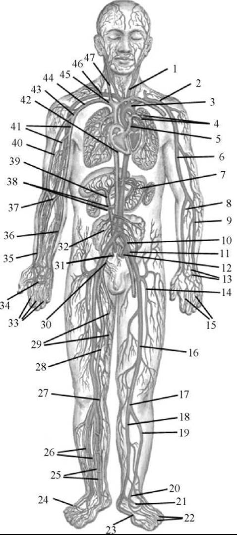
Fig. 201. Circulatory system (General Overview)
I — common carotid artery; 2 — Subclavian Artery; 3 — aortic arch; 4 — pulmonary arteries; 5 — pulmonary trunk; 6 — brachial artery; 7 — left gastric artery and Veins; 8 — radial artery; 9 — ulnar artery; 10 — common iliac artery;
II — external iliac artery; 12 — internal iliac artery; 13 — dorsal carpal arterial network; 14 — deep femoral artery; 15 — dorsal digital Arteries of the hand; 16 — femoral artery; 17 — popliteal artery; 18 — posterior tibial artery; 19 — anterior tibial artery; 20 — dorsalis pedis artery; 21 — lateral plantar artery; 22 — dorsal digital arteries of the FOOT; 23 — medial plantar artery; 24 — plantar venous arch; 25 — posterior tibial veins; 26 — anterior tibial veins; 27 — popliteal vein; 28 — femoral vein; 29 — great saphenous vein; 30 — external iliac vein; 31 — internal iliac vein; 32 — testicular vein; 33 — proper palmar digital arteries and veins; 34 — superficial palmar arterial arch; 35 — radial vein; 36 — ulnar vein; 37 — median cubital vein; 38 — right renal artery and vein; 39 — portal vein; 40 — basilic vein of the upper limb; 41 — brachial veins; 42 — INFERIOR VENA CAVA; 43 — axillary vein; 44 — subclavian vein; 45 — right brachiocephalic vein; 46 — External Jugular Vein; 47 — Internal jugular vein
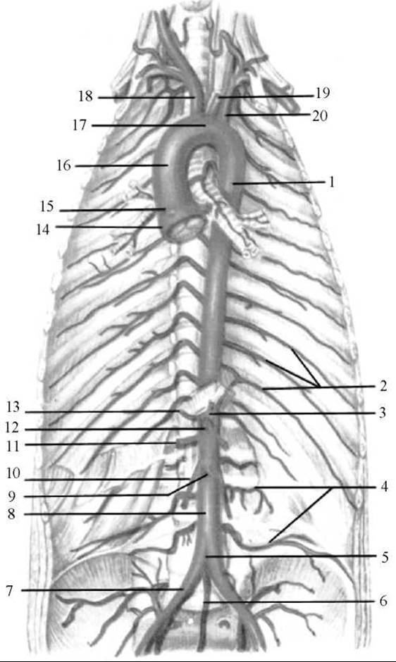
Fig. 202. The aorta and its branches
1 — Thoracic Aorta; 2 — posterior intercostal arteries; 3 — celiac trunk; 4 — lumbar arteries; 5 — aortic bifurcation; 6 — median sacral artery; 7 — right common iliac artery; 8 — Abdominal Aorta; 9 — inferior mesenteric artery; 10 — right testicular artery; 11 — right renal artery; 12 — superior mesenteric artery; 13 — right inferior phrenic artery; 14 — aortic bulb; 15 — right coronary artery; 16 — ascending aorta; 17 — aortic arch; 18 — brachiocephalic trunk; 19 — left common carotid artery; 20 — left subclavian artery
The descending aorta (pars descendens aortae) is its longest section, extending from the level of the 4th thoracic vertebra to the 4th lumbar vertebra, where the aorta bifurcates into the right and left common iliac arteries. The descending aorta is divided into two parts: the thoracic and abdominal parts.
The thoracic aorta (pars thoracica aortae) is located in the thoracic cavity within the posterior Mediastinum. In its superior course, it initially runs anteriorly and to the left of the oesophagus, then at the level of the 8th–4th thoracic vertebrae, it curves around the left side of the oesophagus and passes onto its posterior surface. The azygos vein and Thoracic duct lie to the right of the thoracic aorta, while the parietal Pleura abuts it on the left. The Branches of the thoracic aorta supply the Organs of the thoracic cavity and the chest wall.
The abdominal aorta (pars abdominalis aortae) is the continuation of the thoracic aorta. It begins at the level of the 12th thoracic vertebra where it passes through the aortic hiatus of the Diaphragm, and extends to the midpoint of the body of the 4th lumbar vertebra. At the level of the fourth lumbar vertebra, the abdominal aorta divides into the right and left common iliac arteries. This junction point is termed the aortic bifurcation (bifurcatio aortae). The direct continuation of the abdominal aorta is the median sacral artery, which descends into The Pelvis as a slender, inconstant trunk (homologous to the tail artery in mammals). The abdominal aorta is retroperitoneal, lying on the anterior surface of the lumbar vertebral bodies, slightly to the left of the midline. The inferior vena cava lies to its right, while the Pancreas, the horizontal (inferior) part of the duodenum, and the ROOT of the mesentery of the Small Intestine are situated anteriorly. The abdominal aorta gives rise to branches that supply the abdominal organs and abdominal wall.
Last update: 08/08/2026
Editorial and Educational Adaptation: This material has been compiled based on the primary/original source text. The project team performed an editorial review, corrected technical inaccuracies, structured sections, and adapted the content for an educational format.
What was processed:
- elimination of formatting defects (OCR errors, structural breaks, corrupted characters);
- editorial organization of content;
- standardization of terminology in accordance with academic sources;
- verification of factual statements against the original source text.
All mentions of the author, publication year, and origin of the primary text have been preserved in accordance with the source.