Human Anatomy Part 2 - K. A. Dyubenko A. K. Kolomiyytsev Yu. B. Chaykovsky 2008
Special Part
Cardiovascular system, systema cardiovasculare - Special Part
Blood vessels of the systemic circulation - Arteries of the head and neck - External carotid artery
The External Carotid Artery, a. carotis externa (Fig. 23), supplies Blood to the HEAD and Neck structures. It ascends, passing through the parenchyma of the parotid gland, and lies deeper than the Hypoglossal nerve, the posterior belly of the digastric Muscle (m. digastricus), and the stylohyoid muscle (m. stylohyoideus). Upon reaching the neck of the mandibular articular process, it divides into three groups of branches: anterior, middle, and posterior.
Class="center">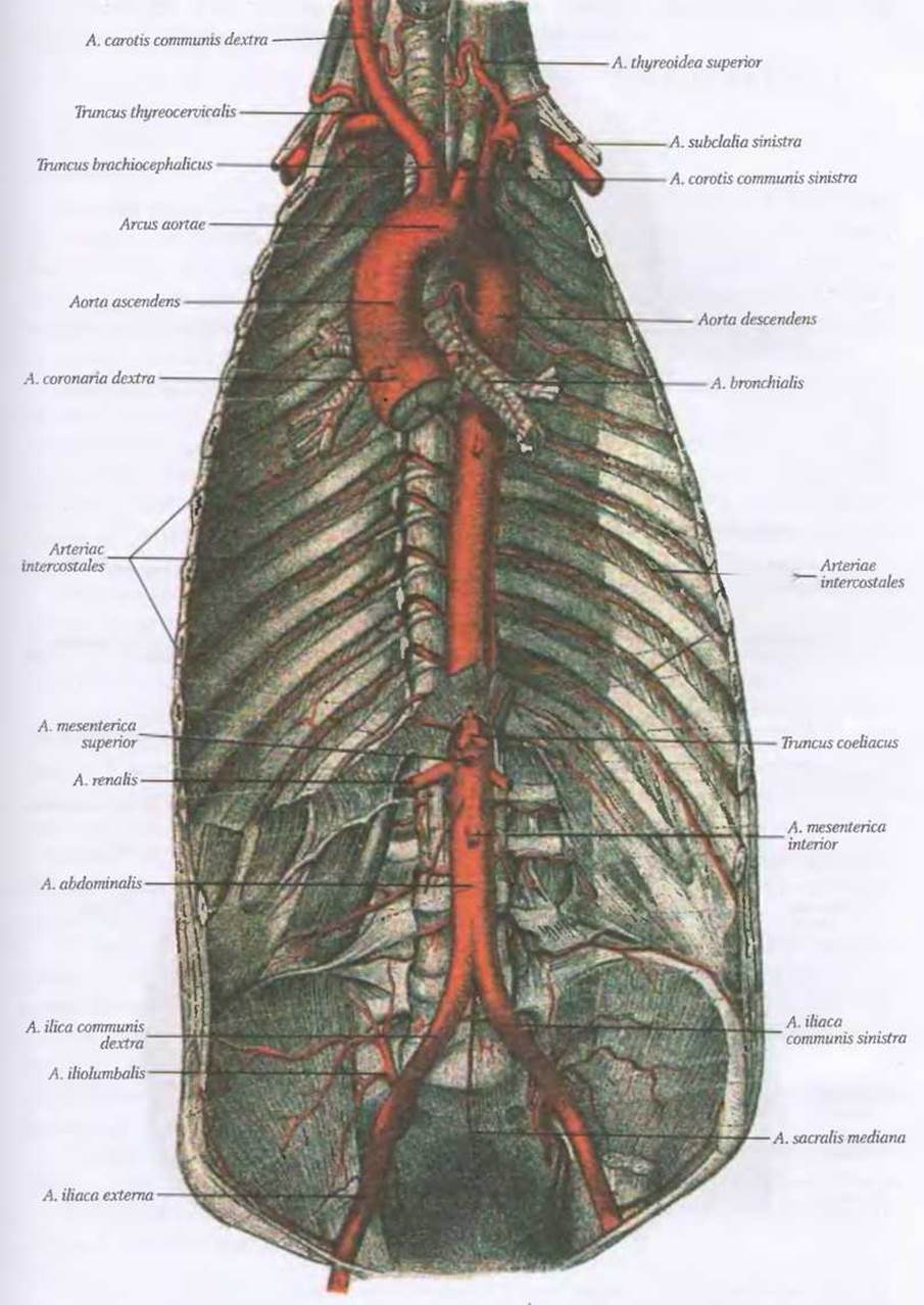
Fig. 21. Aorta and its branches (after Told)
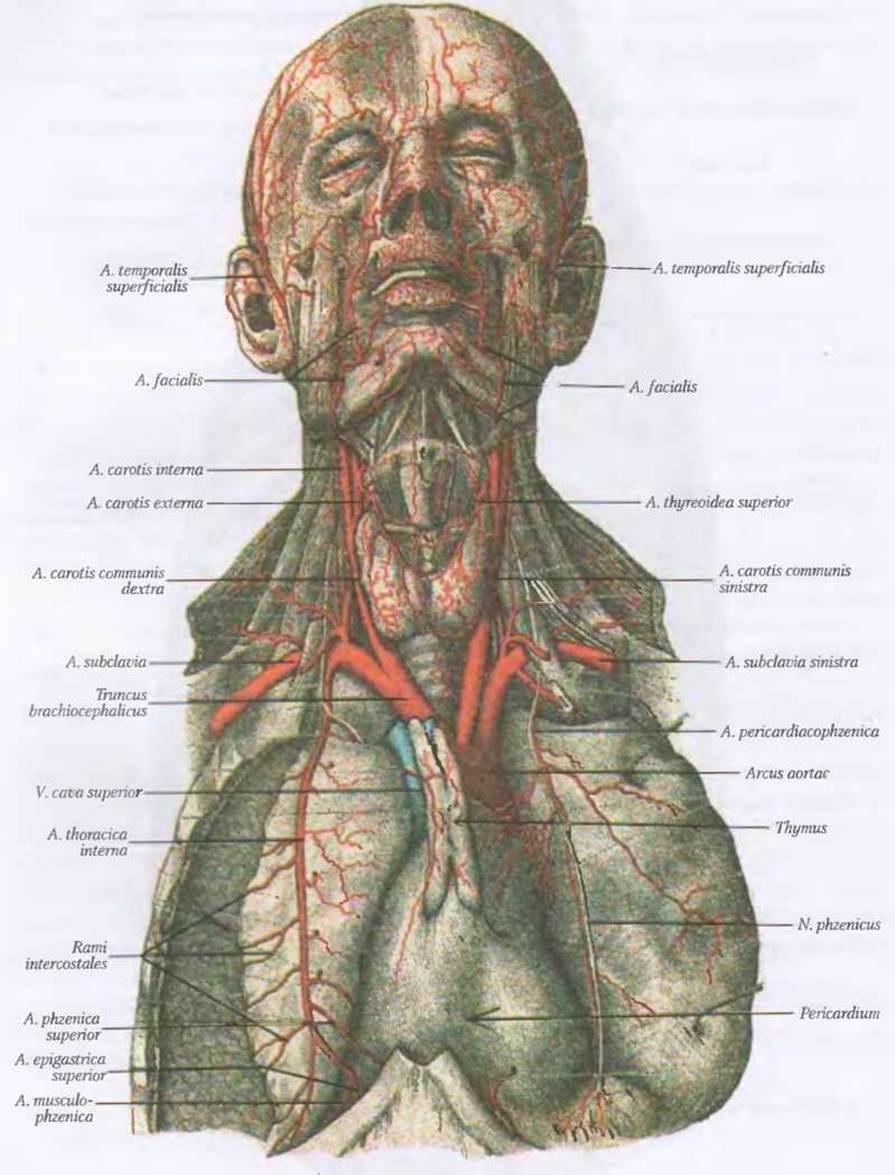
Fig. 22. Arteries of the head, neck, and partially the thoracic cavity (after Told)
Anterior Group of Branches of the external carotid artery:
1. A. thyroidea superior;
2. A. lingualis;
3. A. facialis.
The superior thyroid artery, a. thyreoidea superior (Figs. 22, 25), is a paired vessel that originates from the external carotid artery at the level of the upper border of the thyroid Cartilage, and then runs downward and forward toward The Thyroid Gland. Along its course, it gives off branches to the Hyoid bone and the Larynx. Its largest branch is the superior laryngeal artery, a. laryngea superior, which pierces the thyrohyoid membrane (membrana thyroidea) to supply the mucosa and Muscles of the larynx.
The lingual artery, a. lingualis, is a paired vessel originating from the external carotid artery at the level of the greater horns of the hyoid bone (see Fig. 24). The deep lingual artery, a. profunda linguae, is projected within Pirogov's triangle and penetrates the ROOT of the Tongue, where it anastomoses with the artery of the opposite side. Along its course, it gives off branches to the hyoid bone, the root of the tongue, the palatine Tonsils, and the sublingual gland, continuing toward the tip of the tongue as the a. profunda linguae, where it anastomoses with the contralateral artery.
The facial artery, a. facialis, is a paired vessel originating from the external carotid artery at the level of the angle of the Mandible (Figs. 22, 23). It then courses forward and upward, passing beneath the stylohyoid muscle (m. stylohyoideus), the posterior belly of the digastric muscle (m. digastricus), and the hyoglossus muscle (m. hyoglossus) to reach the submandibular triangle, where it pierces the submandibular gland*.
Near the anterior margin of the masseter muscle, the artery curves around the mandible, emerges onto the face, and reaches the angle of the Mouth. From the angle of the mouth, it extends toward the medial angle of the eye, where its terminal branch—the angular artery, a. angularis—anastomoses with the dorsal nasal artery (a. dorsalis nasi, a branch of the ophthalmic artery). Through this intersystemic anastomosis, during inflammatory conditions of the face (such as Phlegmon), infection can spread into the cranial cavity and cause inflammation of the Meninges (meningitis), a factor that must be considered in clinical practice. Along its course, the facial artery gives off several branches that supply the structures of the facial Skeleton:
1. Ascending palatine artery, a. palatina ascendens – supplies the muscles and mucosa of the Pharynx, the superior pharyngeal constrictor, and the palatine tonsil;
2. Tonsillar branch, ramus tonsillaris – supplies the palatine tonsil;
3. Submandibular branches, rami submandibulares – supply the submandibular gland and anastomose with branches of the a. lingualis;
4. Submental artery, a. submentalis – reaches the chin and supplies the muscles located above the hyoid bone;
5. Inferior labial artery, a. labialis inferior – supplies the lower lip and anastomoses with the contralateral artery;
6. Superior labial artery, a. labialis superior – supplies the upper lip and anastomoses with the corresponding artery of the opposite side.
* The facial artery is injured during tonsillectomy more frequently than other vessels. This is because, within the submandibular triangle, the facial artery forms a loop as it passes along the posterior pole of the submandibular gland near the pharyngeal wall (V. V. Kovanov, 1978).
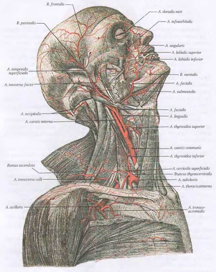
Fig. 23. Arteries of the head and neck according to Told (right lateral view)
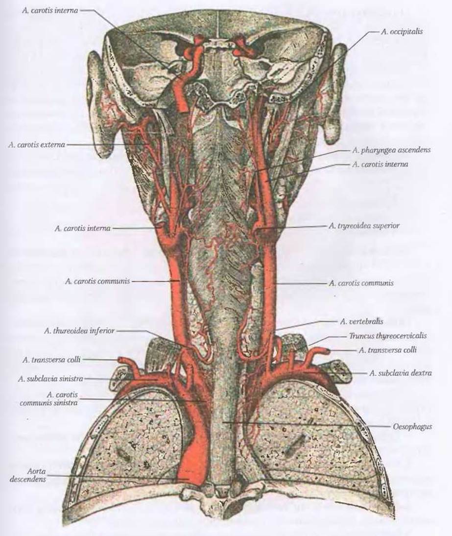
Fig. 23A. Arteries of the head and neck (posterior view) according to Told.
Posterior group of branches of the external carotid artery:
1 A.occipitalis;
2. A.auricularis posterior;
3. Rr.stemocleidomastoidei.
The occipital artery, a.occipitalis (Fig. 23 A), runs towards the mastoid process, where it lies in the groove of the same name and emerges into the occipital region beneath the m.epicrarius, supplying the muscles and Skin of the occipital region, the auricle, and the dura mater of the posterior cranial fossa, which it enters through the jugular foramen.
The posterior auricular artery, a.auricularis posterior (Fig. 23 A), branches off from the external carotid artery superior to the occipital artery*, and courses towards the styloid process, lodging between the external acoustic meatus and the mastoid process of the Temporal bone. The artery branches out in the occipital region, supplying the muscles, skin, and auricle, and also gives off branches to supply the Facial Nerve and the mucous membrane of the tympanic cavity.
* In 30% of cases, it originates as a common trunk with the occipital artery.
The sternocleidomastoid branches, rr.stemocleidomastoidei, arise from the occipital and posterior auricular arteries and supply the muscle of the same name.
Middle group of branches of the external carotid artery:
1. A.pharyngea ascendens;
2. A.temporalis superficialis;
3. A.maxillaris.
The ascending pharyngeal artery, a.pharyngea ascendens, originates from the external carotid artery at the same level as the a.lingualis, runs superiorly along the pharyngeal wall, and its terminal branch reaches the Skull base. The ascending pharyngeal artery supplies the pharynx, soft palate, and dura mater of the posterior cranial fossa.
The superficial temporal artery, a.temporalis superficialis, is a continuation of the external carotid artery trunk (Fig. 23). It runs subcutaneously over the temporalis muscle, anterior to the external acoustic meatus, and reaches the temporal region, where it divides into two terminal branches: ramus frontalis and ramus parietalis. Along its course, it gives off several branches:
1. The transverse facial artery, a.transversa faciei, branches off at the Water/144.html">Origin of the temporal artery, runs inferior to the zygomatic arch, and anastomoses with branches of the a.facialis and a.maxillaris.
2. The parotid branches, ramі parotidei, consist of 2–3 small branches that ramify among the lobules of the parotid gland.
3. The middle temporal artery, a.temporalis media, originates at the level of the root of the zygomatic process of the temporal bone, passes through the fascia temporalis, and supplies the temporalis muscle.
4. The anterior auricular branches, rami auriculares, numbering 3–5 small branches, supply the auricle and the external acoustic meatus.
5. The zygomaticoorbital artery, azygomaticoorbitalis, branches off superior to the external acoustic meatus and runs towards the lateral angle of the eye, where it anastomoses with branches of the a.ophtalmica.
6. The frontal branch, ramus frontalis, is a terminal branch of the a.temporolis superficialis, runs into the regio frontalis, where it branches and anastomoses with branches of the a.ophtalmica.
7. The parietal branch, ramus parietalis, is a terminal branch of the a.temporalis superficialis, reaches the regio ocipitalis, where it anastomoses with branches of the a.occipitalis.
The maxillary artery, a.maxillaris (Fig. 24), is the terminal branch of the external carotid artery. It has a complex course and numerous branches. Topographically and anatomically, its trunk is divided into three segments: the first curves around the neck of the mandible, the second is located in the infratemporal fossa between the lateral surface of the lateral pterygoid and temporalis muscles, and the third lies in the fossa pterygopalatina.
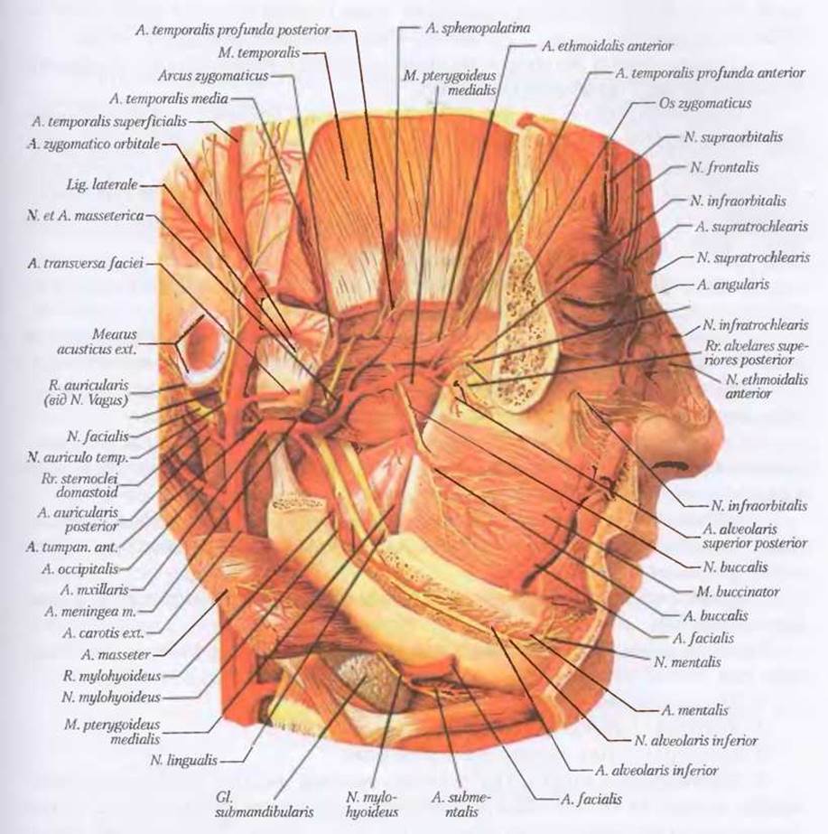
Fig. 24. Maxillary artery, a.maxillaris (after Sobotta)
In the first segment, the maxillary artery gives off the following branches (Fig. 24):
1. The inferior alveolar artery, a.alveolaris inferioris;
2. Deep auricular artery, a.auricularis profunda;
3. Anterior tympanic artery, a.tympanica anterior;
4. Middle meningeal artery, a.meningea media.
1. Inferior alveolar artery, a.alveolaris inferioris, runs between the medial pterygoid muscle and the ramus of the mandible, and then enters the canalis mandibularis. Before entering the canal, the mylohyoid branch, a.mylohyoidea, arises from the inferior alveolar artery to supply the m.mylohyoideus. As it courses through the canal, the inferior alveolar artery gives off branches to the Teeth and gingiva, and its terminal branch, the a.mentalis, emerges through the foramen mentale to supply the chin muscles and skin.
2. Deep auricular artery, a.auricularis profunda, supplies the external acoustic meatus and tympanic membrane.
3. Anterior tympanic artery, a.tympanica anterior, passes through the petrotympanic fissure into the tympanic cavity, where it supplies its mucous membrane.
4. Middle meningeal artery, a.meningea media, enters the middle cranial fossa through the foramen spinosum, runs in the groove of the squamous part of the temporal and parietal bones, and supplies the dura mater and the trigeminal ganglion. It is the largest branch of the first part.
The following branches arise from the second part of the maxillary artery (Fig. 24):
1. Deep temporal arteries, anterior and posterior, aa.temporales profundae anterior et posterior, ascend along the edges of the temporal muscle and branch within it.
2. Masseteric artery, a.masseterica, runs laterally and inferiorly, passes through the mandibular notch (incisura mandibulare), reaches the m.masseter, and branches there.
3. Posterior superior alveolar artery, a.alveolaris superior posterior, its branches pass through the alveolar foramina in the maxillary tuberosity. The artery supplies the teeth, gingiva, and the mucous membrane of the maxillary sinus (sinus maxillaris).
4. Buccal artery, a.buccalis, runs forward and downward, reaches the m.buccinator where it branches; it supplies the cheek and the maxillary gingiva, anastomosing with branches of the a.facialis.
5. Pterygoid branches, rami pterygoidei, numbering 3-4, supply the pterygoid muscles.
The third part of the maxillary artery enters the pterygopalatine fossa (fossa pterygopalatina), giving rise to the following arteries (Fig. 24):
1. Infraorbital artery, a.infraorbitalis;
2. Descending palatine artery, a.palatina descendens;
3. Sphenopalatine artery, a.sphenopalatina.
1. Infraorbital artery, a.infraorbitalis, enters the Orbit through the inferior orbital fissure, lies in the infraorbital groove (sulcus infraorbitalis), then in the infraorbital canal (canalis infraorbitalis), and emerges onto the anterior surface of the Maxilla through the corresponding foramen. Within the orbital groove or canal, the artery gives off the anterior superior alveolar arteries, aa.alveolares superiores anteriores, which supply the upper anterior teeth, gingiva, and the mucous membrane of the sinus maxillaris. Passing through the orbit, the a.infraorbitalis gives off branches to the extraocular muscles. Upon emerging onto the face, the a.infraorbitalis supplies the lower eyelid, lacrimal sac, upper lip, and cheek, and also anastomoses with branches of the a.facialis.
2. Descending palatine artery, a.palatina descendens, passes through the greater palatine canal (canalis palatinus major) and reaches the Hard and Soft palate via two branches, the greater and lesser palatine arteries (a.palatina major et minor), supplying the mucous membrane of the palate and upper gingiva. The a.canalis pterygoidei arises from the descending palatine artery to supply the nasopharynx and the auditory tube.
3. Sphenopalatine artery, a.sphenopalatina, enters the Nasal cavity through the sphenopalatine foramen, where it branches and supplies the nasal mucosa (lateral wall and septum).
Last update: 08/08/2026
Editorial and Educational Adaptation: This material has been compiled based on the primary/original source text. The project team performed an editorial review, corrected technical inaccuracies, structured sections, and adapted the content for an educational format.
What was processed:
- elimination of formatting defects (OCR errors, structural breaks, corrupted characters);
- editorial organization of content;
- standardization of terminology in accordance with academic sources;
- verification of factual statements against the original source text.
All mentions of the author, publication year, and origin of the primary text have been preserved in accordance with the source.