Human Anatomy Part 1 - K. A. Dyubenko, A. K. Kolomiysev, Yu. B. Chaykovsky 2002
General Part
Structure of the Human Body
Tissues - Nervous Tissue - Neuroglia
Neuroglia is a supportive type of Nervous Tissue that performs vital Functions. The Classification of neuroglial types can be schematically represented as follows:
Class="center">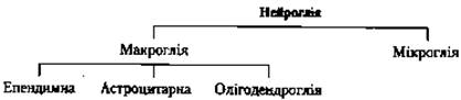

Fig. 11. Structure of a neuron (A) and ultrastructural Organization of a nerve fiber at various levels (B) (after T. N. Radostina, Yu. I. Afanasyev, and L. S. Rumyantsev):
1 - dendrites; 2 - neuron Cell body; 3 - axon; 4 - node of Ranvier; 5 - nerve ending; 6 - Nucleus of a Schwann cell; 7 - axon cylinder; 8 - nucleus of a neuroleimocyte; 9 - basement membrane; 10 - Mitochondria; 11 - neurofilaments; 12 - myelin sheath
Ependymal Glia lines the cavities of the Brain (the ventricles and the central canal of the Spinal Cord) and its choroid plexuses. In these cavities, it is represented by cuboidal or prismatic Cells bearing one or several motile cilia on their apical surface. Long processes emerge from the basal part of ependymocytes and penetrate into the brain tissue. The ependymal cells covering the outer surface of the plexus Blood Vessels are cuboidal in shape. Together with the vascular wall, they participate in The formation of the CEREBROSPINAL FLUID.
Astroglial glia is formed by stellate (branched) cells. There are two MAIN TYPES OF astrocytes: protoplasmic (short-branched) astrocytes, which have short processes containing numerous glial filaments formed by Fibrillar Proteins in their Cytoplasm, and fibrous (long-branched) astrocytes, which contain fewer filaments assembled into bundles. Protoplasmic astrocytes predominate in the Gray matter, whereas fibrous ones predominate in the White matter OF the brain. Some astrocytic processes (perivascular feet) extend toward blood capillaries and, spreading out on their outer surface, form glial limiting membranes that, together with the Capillary Wall, constitute an essential component of the blood-brain barrier.
Oligodendroglia comprises a numerous group of cells adjacent to neuronal cell bodies or their processes. Establishing connections with neurites during the Embryogenesis of nervous tissue, oligodendroglia participates in the formation of nerve fiber sheaths. These cells are characterized by a poorly developed network of glial filaments, yet they contain a high Abundance of general-purpose Organelles and trophic inclusions.
Microglia consists of small, branched cells capable of active phagocytosis. Some authors consider these cells to be macrophages of the Central Nervous system originating from mesenchyme during embryogenesis, whereas others view them as immature astrocytes capable of proliferation and transformation into astrocytes.
The Functional Significance of neuroglia (primarily astroglial) lies in its supportive and mechanical role. In addition, gliocytes provide reliable electrical insulation for neuronal cell bodies, their processes, and synapses, thereby preventing electrical crosstalk between neurocytes during the Propagation of Excitation. Oligodendroglial cells take an active part in the trophic support of neurocytes and the formation of nerve fiber sheaths, while microgliocytes (CNS macrophages) perform a protective function.
Last update: 08/08/2026
Editorial and Educational Adaptation: This material has been compiled based on the primary/original source text. The project team performed an editorial review, corrected technical inaccuracies, structured sections, and adapted the content for an educational format.
What was processed:
- elimination of formatting defects (OCR errors, structural breaks, corrupted characters);
- editorial organization of content;
- standardization of terminology in accordance with academic sources;
- verification of factual statements against the original source text.
All mentions of the author, publication year, and origin of the primary text have been preserved in accordance with the source.