HUMAN MEDICAL BIOLOGY, ANATOMY, PHYSIOLOGY, AND PATHOLOGY - Ya.I. Fedonyuk 2010
BIOLOGY
CHAPTER 1. BIOLOGICAL FOUNDATIONS OF HUMAN VITAL ACTIVITY
1.3. MOLECULAR-GENETIC AND CELLULAR LEVELS OF LIFE ORGANIZATION
1.3.3. The Hereditary Apparatus of Eukaryotic Cells and Its Functioning: The Molecular Level
DNA: Chemical Composition, Spatial Organization, and Role in the Selection/27.html">Realization of Genetic Information
DNA (deoxyribonucleic acid) is a high-molecular-weight organic compound, a biopolymer whose monomers are NUCLEOTIDES linked together into a long polynucleotide chain. The relative molecular mass of DNA reaches 1,500,000–2,000,000 or more. The number and arrangement order of nucleotides in it constitute the Introduction/19.html">Primary Structure of the DNA molecule. Each nucleotide consists of three components: 1) a phosphoric acid residue; 2) a pentose sugar, deoxyribose (hence the name of this acid: deoxyribonucleic); 3) one of four nitrogenous bases: adenine (A), guanine (G), cytosine (C), and thymine (T). Adenine and guanine belong to the Class of Purines, while cytosine and thymine belong to the class of Pyrimidines. Within the nucleotide, deoxyribose is located in the middle—between the phosphoric acid and the nitrogenous base. A compound formed by a nitrogenous base and a pentose (deoxyribose) is called a nucleoside (deoxyribonucleoside). Nucleotides are linked to each other by phosphodiester bonds formed between the 3' and 5' hydroxyl groups of the deoxyribose sugars of adjacent nucleotides. Two ends are distinguished in a DNA polynucleotide chain. One end bears a hydroxyl group (-OH) attached to the 3' carbon of the deoxyribose sugar, while the other end contains a phosphoric acid residue at the 5' position of the sugar.
DNA is characterized by certain regularities established in 1950 by the American biochemist E. Chargaff (born in Chernivtsi). Chargaff's rules state that:
1) the sum of purine bases is equal to the sum of pyrimidine bases: A+G = T+C;
2) The amount of bases with a keto group in the 6th position is equal to the amount of bases with an amino group in the 6th position: G+T = A+C, or (G+T)/(A+C) = 1;
3) the content of adenine equals the content of thymine, and the content of guanine equals the content of cytosine (the rule of equivalence): A=T, G=C. According to Chargaff's rules, the DNA composition of different organisms can vary only in the (A+T)/(G+C) ratio, but for organisms of a given species, this ratio is constant: in humans it is 1.54, and in the bacterium *Escherichia coli*, 1.0.
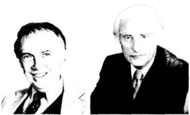
James Watson, Francis Crick
(born 1928). (born 1916).
In 1953, the American biochemist J. Watson and the English physicist F. Crick, working at Cambridge University, proposed a model of the Spatial Structure of DNA AS A double helix based on chemical and X-Ray Diffraction data (Nobel Prize, 1962). According to the Watson-Crick model, a DNA molecule consists of two polynucleotide chains wound to the right around a common axis into a double helix. The two chains in the DNA molecule are antiparallel: one chain runs in the 5' → 3' direction, and the other in the 3' → 5' direction. The DNA double helix is maintained by Hydrogen Bonds between the bases of opposite chains. Base pairing is carried out according to THE PRINCIPLE OF complementarity: adenine of one chain is always paired with thymine of the other (A-T), and guanine with cytosine (G-C). Two hydrogen bonds form between adenine and thymine, and three between guanine and cytosine. The paired bases are located between the two chains, perpendicular to the central axis. The entire structure resembles a spiral staircase, the sides of which are formed by the sugar-phosphate backbone, and the steps by the paired bases. The diameter of the helix is 2 nm, the distance between nitrogenous bases along the axis of the helix is 0.34 nm, and ten Base Pairs form one complete turn of The Double Helix with a length of 3.4 nm (Figs. 1.38, 1.39).
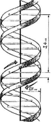
Fig. 1.38. Diagram of The structure of the DNA double helix (bihelix).
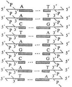
Fig. 1.39. Diagram illustrating the complementarity (A-T, G-C) and antiparallelism of the two strands of a DNA molecule.
Different forms of the DNA double helix exist (A, B, C, D, Z). The B-form is described above. In a living Cell, the double helix, which constitutes the Secondary structure of DNA, does not exist as an unraveled thread, but is additionally coiled in space with the help of histone Proteins, forming a superhelix—the tertiary structure. Supercoiling ensures the compact packing of the giant DNA molecule within the chromosome. In addition to nuclear DNA, Eukaryotic Cells contain Mitochondrial DNA (in plants and animals) and plastid DNA (in plants). Mitochondrial DNAs are significantly smaller than nuclear ones, have a double-stranded circular structure, and are not bound to Histones.
DNA molecules in various plants and animals are constructed from the same four types of nucleotides: adenylic, guanylic, cytidylic, and thymidylic (A, G, C, T), yet they differ in both the quantity and sequence order of these nucleotides. This circumstance determines the universality and, at the same time, the uniqueness of DNA. The DNA of each species is characterized by relative stability and species Specificity. The principle of complementarity is a crucial feature of the DNA helix structure that enables it to perform two Functions: 1) the autocatalytic (autosynthetic) function—DNA Synthesis on a DNA template (Replication), and 2) the heterocatalytic (heterosynthetic) function—RNA Synthesis on a DNA template (METABOLISM/31.html">Transcription). Both functions are related to the Biological Role of DNA as the carrier of Genetic information: the storage, transmission, and realization of genetic information.
Last update: 08/08/2026
Editorial and Educational Adaptation: This material has been compiled based on the primary/original source text. The project team performed an editorial review, corrected technical inaccuracies, structured sections, and adapted the content for an educational format.
What was processed:
- elimination of formatting defects (OCR errors, structural breaks, corrupted characters);
- editorial organization of content;
- standardization of terminology in accordance with academic sources;
- verification of factual statements against the original source text.
All mentions of the author, publication year, and origin of the primary text have been preserved in accordance with the source.