ATLAS OF HUMAN ANATOMY - G.L. Bilich - 2014
Muscular System
MUSCLES OF THE PPECTORAL GIRDLE
Muscles of the Free Upper Limb
Muscles of the Arm
The Muscles of the arm are divided into two groups: anterior and posterior. The anterior muscles are flexors (coracobrachialis, biceps brachii, and brachialis), and the posterior ones are extensors (triceps brachii and anconeus) (see Figs. 303, 304, 305). The anterior group is separated from the posterior by Cytology/practical/45.html">Dense Connective Tissue medial and lateral intermuscular septa (septum intermusculare brachii mediale and septum intermusculare brachii laterale), which fuse with the periosteum of the humerus and form a soft connective tissue Skeleton of the arm. The medial intermuscular septum of the arm is thicker than the lateral one and terminates inferiorly on the medial epicondyle of the humerus.
Anterior Group of arm muscles. The biceps brachii (m. biceps brachii) is a long fusiform Muscle located on the anterior aspect of the arm, having two heads—short and long (Figs. 313, 314). The long HEAD (caput longum) originates from the supraglenoid tubercle of the scapula by a rounded tendon that passes from top to bottom through the capsule of the shoulder joint. Within the joint cavity, the tendon is covered by a synovial membrane. Upon emerging onto the arm
the tendon lies in the intertubercular sulcus, being enveloped by the intertubercular synovial sheath (Vagina synovialis intertubercularis). The short head (caput breve) originates at the apex of the coracoid process of the scapula together with the coracobrachialis muscle. The long and short heads run side by side from top to bottom and, at the level of the middle of the arm, unite into a common fusiform muscle belly. Near the elbow joint or above it, the belly of the biceps continues into a long tendon that attaches to the radial tuberosity.
Class="center">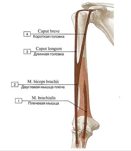
Fig. 313. Origin and insertion sites of the brachialis and biceps brachii muscles (diagram):
1 — Brachialis; 2 — Biceps brachii 3 — Long head; 4 — Short head
Near the tendon insertion site, there is the bicipitoradial bursa (bursa bicipitoradialis). A thin, broad, dense aponeurosis of the biceps brachii—the Pirogov fascia (aponeurosis) (aponeurosis musculi bicipitis brachii)—extends from the anteromedial surface of the tendon; it covers the cubital fossa anteriorly and blends into the forearm fascia on its medial side, anterior to the pronator teres. In shape, this aponeurosis resembles a trapezoid, which is why it is also referred to as the trapezoidal fascia.
Function: the biceps flexes the arm at the shoulder joint; flexes and supinates the forearm at the elbow joint; supinates the pronated forearm (Fig. 315).
Biceps brachii muscle; tendon.
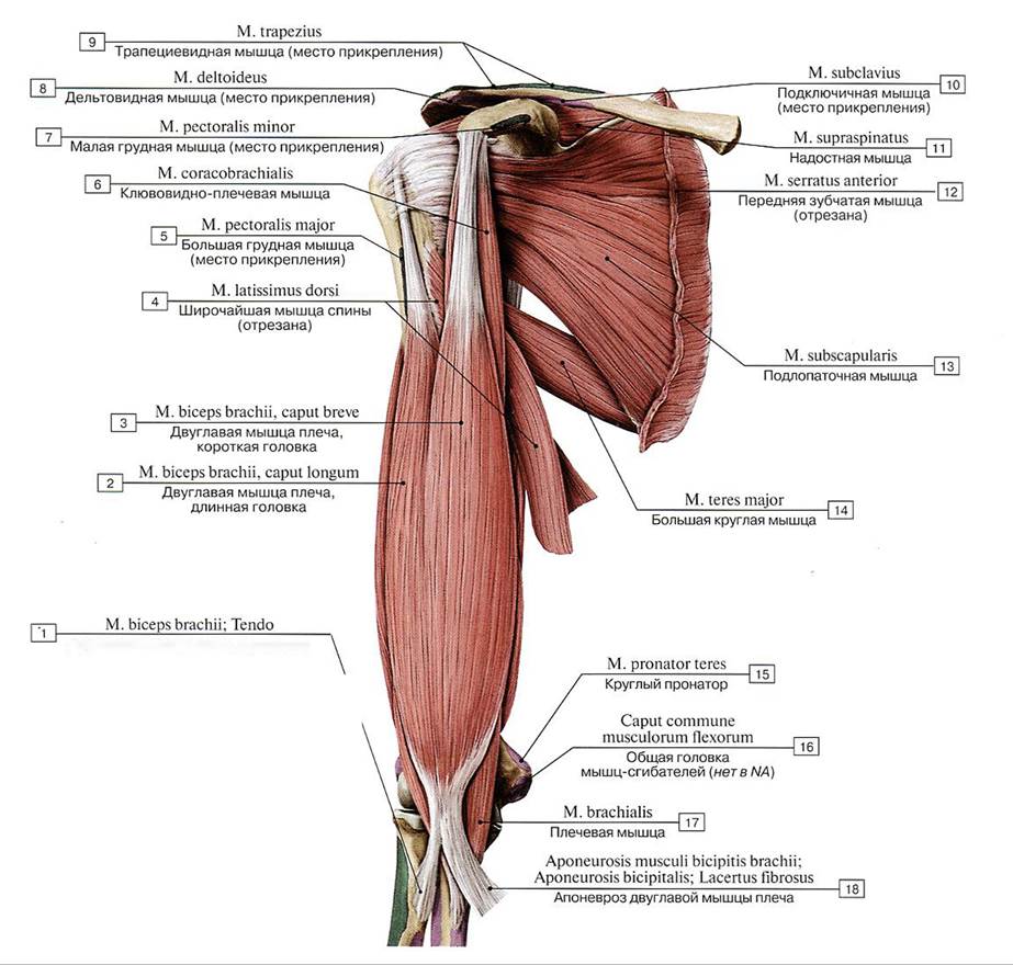
Fig. 314. Subscapularis, teres major, and biceps brachii muscles of the right upper limb, anterior view:
1 — Biceps brachii; Tendon; 2 — Biceps brachii, long head; 3 — Biceps brachii, short head; 4 — Latissimus dorsi; 5 — Pectoralis major; 6 — Coracobrachialis; 7 — Pectoralis minor; 8 —- Deltoid; 9 — Trapezius; 10 — Subclavius; 11 — Supraspinatus; 12 — Serratus anterior; 13 — Subscapularis; 14 — Teres major; 15 — Pronator teres; 16 — Caput commune of flexors; 17 — Brachialis; 18 — Bicipital aponeurosis
Blood supply: axillary, brachial, superior and inferior ulnar collateral, and radial recurrent Arteries.
Innervation: musculocutaneous nerve (CV—CVIII]
Coracobrachialis muscle (m. coracobrachialis — Casserio's muscle — appears as a flattened cord located medial to the short head of the biceps brachii, with whose tendon this muscle is fused (Giulio Casserio, 1545–1616, Italian anatomist). The muscle originates at the apex of the coracoid process, runs from top to bottom, and attaches via a broad flat tendon to the anterior and medial surfaces of the humerus, extending from the crest of the lesser tubercle down to the middle of this bone (Fig. 316). Some muscle fascicles blend into the medial intermuscular septum of the arm. Within the substance of the coracobrachialis muscle, There is a narrow cleft through which the musculocutaneous nerve supplying this muscle (and adjacent ones) passes.

Fig. 315. Functions of the biceps brachii muscle (diagram):
1 — Biceps brachii; 2 — Biceps brachii; Tendon; 3 — Radial tuberosity; 4 — Ulna; 5 — Radius
Supination of the forearm by the biceps brachii with the forearm flexed.
A. The forearm is pronated and flexed at the elbow joint (right hand, medial view)
B. Cross-section at the level of the radial tuberosity, with the forearm pronated (proximal view).
C. Forearm supinated and flexed at the elbow joint (right arm, medial view)
D. Cross-section at the level of the radial tuberosity, with the forearm supinated (proximal view).
When the forearm is flexed, the biceps brachii acts as a powerful supinator In addition to its function as a flexor, because the arm in this position is almost perpendicular to the axis of pronation/supination. This explains why supination is particularly effective when the forearm is flexed. When the forearm is pronated (A), the tendon of the biceps brachii is, so to speak, wrapped around the radius. When the muscle flexes the forearm from this position, the tendon unwraps (B).
Between the humerus on the one hand, and the coracobrachialis, the short head of the biceps brachii, and the distal tendon of the subscapularis on the other, there lies the coracobrachial bursa (bursa coracobrachialis).
Function: the coracobrachialis flexes the arm at the shoulder joint and adducts it toward the trunk, rotates the pronated arm outward, and, with a fixed arm, pulls the scapula forward and downward.
Blood supply: anterior and posterior circumflex humeral arteries.
Innervation: musculocutaneous nerve (CV—CVII).
The brachialis (m. brachialis) is a wide, flat muscle located in the lower half of the anterior region of the arm, covered by the biceps. The brachialis originates by muscular bundles from the lower two-thirds of the shaft of the humerus distal to the deltoid tuberosity, as well as from the medial and lateral intermuscular septa of the arm. The Water/144.html">Origin of the brachialis embraces the insertion site of the deltoid muscle with two bellies. The brachialis runs downward and inserts into the tuberosity of the ulna (see Fig. 313, Fig. 316). Bundles of the deep portion of the muscle tendon weave into the capsule of the elbow joint. The fibers of the brachialis that diverge from it and terminate in the anterior part of the capsule of the elbow joint are termed the articular muscle of the elbow or Portal's muscle (m. capsularis subbrachialis) (Antoine Portal, 1742–1832, French anatomist).
Function: flexes the forearm at the elbow joint.
Blood supply: superior and inferior ulnar collateral arteries, deep artery of the arm, radial recurrent artery.
Innervation: musculocutaneous nerve (CV—CVII).
Posterior muscle group of the arm. The triceps brachii (m. triceps brachii) is a thick, fusiform muscle occupying the posterior aspect of the arm along its entire length. The muscle has three heads: long, lateral, and medial (Fig. 317, see Figs. 304, 305). The long head (caput longum) originates by a thick, short, rounded tendon from the infraglenoid tubercle of the scapula. The muscle belly of the long head first runs downward between the teres minor and teres major muscles and then proceeds to the middle of the posterior aspect of the arm, where it joins with the bundles of the lateral and medial heads. Often, the tendon is connected by a narrow slip to the tendon of the latissimus dorsi.

Fig. 316. Teres major, coracobrachialis, and brachialis muscles, right, anterior view:
1 — Deltoid; 2 — Pectoralis major; 3 — Biceps brachii, long head; 4 — Latissimus dorsi; 5 — Biceps brachii, short head; 6 — Supraspinatus; 7 — Subscapularis; 8 — Trapezius; 9 — Pectoralis minor; 10 — Subclavius; 11 — Serratus anterior; 12 — Subscapularis; 13 — Teres major; 14— Coracobrachialis; 15— Brachialis; 16 — Pronator teres; 17 — Caput commune of flexors; 18 — Radial tuberosity
The lateral head (caput laterale) originates by tendinous and muscular bundles from the posterolateral surface of the humerus, between the insertion site of the teres minor above and the radial groove below, as well as from the posterior surface of the lateral intermuscular septum. The lateral head is partially covered by the deltoid muscle. The bundles of this head run downward and medially, covering the radial groove, which contains the radial nerve of the same name and the deep Vessels of the arm.
The medial head (caput mediale) is shorter than the lateral head and originates by muscular bundles from the posterior surface of the humerus medial to the radial groove, between the insertion site of the teres major above and the olecranon fossa below, as well as from the medial and lateral intermuscular septa inferior to the radial groove. The greater part of the medial head is covered by the lateral head, with which it partially fuses. Between the origins of the medial and lateral heads and the radial groove lies the narrow humeromuscular canal, through which pass the radial nerve and Blood Vessels.

Fig. 317. Right triceps brachii and anconeus muscles (all other muscles removed), posterior view (A — lateral head partially removed, B — long head partially removed):
1 — Triceps brachii, long head; 2 — Coracoid process; 3 — Acromion; 4 — Greater tubercle; 5 — Infraglenoid tubercle; 6 — Triceps brachii, lateral head; Tendon; 7 — Triceps brachii, long head; Tendon; 8 — Radial groove; Groove for radial nerve; 9 — Triceps brachii, medial head; deep head; 10— Triceps brachii; Tendon; 11 — Medial epicondyle; 12 — Lateral epicondyle; 13 — Olecranon; 14 — Anconeus; 15 — Shaft of humerus; Body of humerus; 16 — Triceps brachii, lateral head
The three heads unite at approximately the middle of the posterior aspect of the arm, forming a common belly that transitions into a flat, broad tendon inserting into the olecranon of the ulna. Some bundles of the triceps brachii blend into the capsule of the elbow joint and the Fascia of the forearm. Beneath the tendon lies the subtendinous bursa of the triceps brachii (bursa subtendinea musculi tricipitis brachii). Within the tendon, near its insertion into the olecranon, there is also an intratendinous olecranon bursa (bursa intratendinea olecrani). These bursae do not communicate with the cavity of the elbow joint.
Function: the triceps brachii extends the forearm at the elbow joint; the long head extends the arm at the shoulder joint and adducts it to the trunk.
Blood supply: posterior circumflex humeral artery, deep artery of the arm, superior and inferior ulnar collateral arteries.
Innervation: radial nerve (CV—CVIII).
The anconeus muscle (m. anconeus), sometimes referred to as the fourth head of the triceps brachii, is a flat, triangular muscle located posteriorly at the elbow joint, with whose capsule it is blended. The anconeus originates from the posterior surface of the lateral epicondyle of the humerus and the radial collateral ligament, running inferiorly and medially. It inserts onto the posterolateral surface of the Base of the olecranon process, the proximal ulna, and the forearm fascia (see Fig. 317).
Function: the anconeus extends the forearm at the elbow joint and pulls the Joint Capsule posteriorly.
Blood supply: recurrent interosseous artery, deep brachial artery.
Innervation: radial nerve (Cv—CVIII).
Last update: 08/08/2026
Editorial and Educational Adaptation: This material has been compiled based on the primary/original source text. The project team performed an editorial review, corrected technical inaccuracies, structured sections, and adapted the content for an educational format.
What was processed:
- elimination of formatting defects (OCR errors, structural breaks, corrupted characters);
- editorial organization of content;
- standardization of terminology in accordance with academic sources;
- verification of factual statements against the original source text.
All mentions of the author, publication year, and origin of the primary text have been preserved in accordance with the source.