ATLAS OF HUMAN ANATOMY - G.L. Bilich - 2014
Muscular System
MUSCLES OF THE SHOULDER GIRDLE
Muscles of the Free Upper Limb
Muscles of the Forearm
The numerous Muscles of the forearm perform essential Functions; most of them act across multiple joints (the elbow, proximal and distal radioulnar, wrist, and the JOINTS OF THE hand and fingers), classifying them as multijarticular muscles. Similar to the arm muscles, the forearm muscles are divided into two groups: anterior and posterior. The anterior group includes seven flexors of the wrist and fingers — the brachioradialis, flexor carpi radialis, flexor carpi ulnaris, palmaris longus (which is sometimes absent), flexor digitorum superficialis, flexor digitorum profundus, and flexor pollicis longus — as well as two pronators: the pronator teres and pronator quadratus. The finger flexors execute exceptionally delicate and highly differentiated movements.
The posterior group (extensors of the wrist and fingers) comprises the following muscles: the extensor carpi radialis longus and brevis, extensor carpi ulnaris, extensor digitorum, extensor digiti minimi, extensor indicis, extensor pollicis longus and brevis, abductor pollicis longus, and one supinator. Both the anterior and posterior forearm muscles are arranged in several layers.
Anterior Group of forearm muscles. The anterior forearm muscles (flexors) are arranged in four layers. The superficial layer (extending from the radius toward the ulna) contains the brachioradialis, pronator teres, flexor carpi radialis, palmaris longus, and flexor carpi ulnaris. The second layer contains the flexor digitorum superficialis. The third layer contains the flexor digitorum profundus (on the ulnar side) and the flexor pollicis longus (on the radial side). The deepest, fourth layer houses the pronator quadratus. Each Muscle is enclosed in its own fascial sheath.
Most muscles on the anterior aspect of the forearm originate from the lateral (radial) and medial (ulna) sides of the humerus, specifically from its epicondyles.
First (superficial) layer of the forearm muscles (Figs. 318, 319). The brachioradialis (m. brachioradialis) is a fusiform muscle located anteriorly and laterally to the brachialis, forming the lateral boundary of the cubital fossa. It originates with muscular bundles from the lateral supracondylar ridge of the humerus and the lateral intermuscular septum of the arm, extending toward the hand. At the middle of the forearm, the muscle belly transitions into a narrow, flat tendon that runs along the lateral border of the forearm to insert onto the lateral surface of the distal end of the radius, just proximal to its styloid process. In its distal course, the tendon passes directly deep to the tendons of the abductor pollicis longus and extensor pollicis brevis.
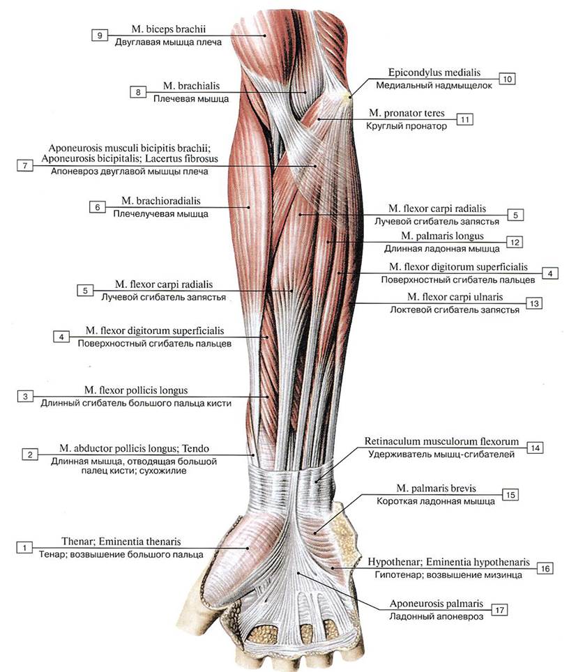
Class="center">Fig. 318. Anterior group of muscles of the right forearm, superficial layer, anterior view:
1 — Thenar eminence; 2 — Abductor pollicis longus; Tendon; 3 — Flexor pollicis longus; 4 — Flexor digitorum superficialis; 5 — Flexor carpi radialis; 6 — Brachioradialis; 7 — Bicipital aponeurosis; 8 — Brachialis; 9 — Biceps brachii; 10 — Medial epicondyle; 11 — Pronator teres; 12 — Palmaris longus; 13 — Flexor carpi ulnaris; 14 — Flexor retinaculum; 15— Palmaris brevis; 16 — Hypothenar eminence; 17— Palmar aponeurosis

Fig. 319. Anterior group of muscles of the right forearm, First and Second layers (A — general view, B — sites of origin and insertion (diagram)):
1 — Base of metacarpal [II]; 2— Flexor digitorum superficialis; 3 — Flexor carpi radialis; 4— Pronator teres; 5 — Radial tuberosity; 6 — Medial epicondyle; Caput commune of flexors; 7— Palmaris longus; 8— Flexor carpi ulnaris; 9— Pisiform; 10 — Hook of hamate; 11 — Base of metacarpal [V]; 12 —Palmar aponeurosis; 13— Middle phalanx [II—V]
Function: The brachioradialis flexes the forearm at the elbow joint, rotates the radius outward, and supinates the hand when the elbow is extended. From a supinated position, this muscle can pronate the hand, which is why it is also known as the regulator of the radius (m. regulator radii).
Blood supply: Radial collateral and recurrent radial Arteries, and the radial artery.
Innervation: Radial nerve (CV—CVIII).
The pronator teres (m. pronator teres) is a short, round muscle located inferior to the elbow joint, deep to the bicipital aponeurosis. Its origin is split into two parts (humeral and ulnar heads), with the median nerve passing between them. The humeral (large) HEAD (caput humerale) originates from the medial epicondyle of the humerus, the medial intermuscular septum of the arm, and the antebrachial fascia. The ulnar (small) head (caput ulnare) originates by a short tendon from the medial margin of the ulnar tuberosity and its coronoid process. Both heads unite, course downward and laterally, and insert via a short, flat tendon at the midpoint of the lateral surface of the radius. The muscle passes anterior to the tendon of the biceps brachii, beneath its aponeurosis, thereby forming the inferomedial boundary of the cubital fossa.
Function: The pronator teres pronates (turns inward) the forearm and flexes it at the elbow joint.
Blood supply: Muscular Branches of the brachial, ulnar, and radial arteries.
Innervation: Median nerve (CV—ThI).
The flexor carpi radialis (m. flexor carpi radialis) is a fusiform muscle originating from the medial epicondyle of the humerus, the fascia, and the medial intermuscular septum of the arm. It lies between the brachioradialis laterally and the palmaris longus medially. At approximately the middle of the forearm, the muscle belly of the flexor carpi radialis transitions into a long, flat tendon that passes beneath the flexor retinaculum through the groove of the trapezium bone (beneath the transverse carpal ligament) and inserts onto the palmar surface of the bases of Metacarpal bones II–III. A synovial bursa, the bursa of the flexor carpi radialis (bursa moscuii flexoris carpi radialis), is situated between the muscle tendon and the scaphoid bone.
A variant muscle in this region may be the short flexor carpi radialis — Wood's muscle (named after John Wood, 1827–1891, an English surgeon). Its shape, size, and position are highly variable. It originates on the lateral or anterior surface of the radius and inserts onto the metacarpal or Carpal Bones. Function: flexion of the hand.
Function: the flexor carpi radialis flexes the hand (wrist) and abducts it (together with the extensor carpi radialis longus), and flexes the forearm at the elbow joint.
Blood supply: muscular branches of the brachial, ulnar, and radial arteries.
Innervation: median nerve (CV—ThI).
The palmaris longus (m. palmaris longus) is a fusiform muscle located superficially on the PALMAR ASPECT OF the forearm. The muscle originates from the medial epicondyle of the humerus, the forearm fascia, and the adjacent intermuscular septa of the forearm via a short muscle belly. At the middle of the forearm, the belly transitions into a long, ribbon-like tendon that crosses onto the hand superficial to the flexor retinaculum and expands into a broad, thick Connective Tissue plate known as the palmar aponeurosis.
Function: the palmaris longus tightens the palmar aponeurosis, and flexes the hand and forearm.
Blood supply: radial artery.
Innervation: median nerve (CV—ThI).
The flexor carpi ulnaris (m. flexor carpi ulnaris) is a flat, triangular, unipennate muscle with two short heads: humeral and ulnar. The humeral head (caput humerale) arises from the medial epicondyle of the humerus and the medial intermuscular septum of the arm. The ulnar head (caput ulnare) originates from the medial margin of the olecranon, the posterior border of the ulna, and the forearm fascia. In the upper third of the forearm, both heads unite to form a common belly that runs downward along the medial side of the forearm. At the middle of the forearm, the belly continues into a long tendon that inserts into the pisiform bone, the hook of the hamate bone, the pisometacarpal and pisohamate ligaments, and the Base of the 5th metacarpal bone. At the site where the tendon attaches to the pisiform bone, there is the bursa of the flexor carpi ulnaris (bursa musculi flexoris carpi ulnaris). The ulnar nerve passes through the cleft between the humeral and ulnar heads.

Fig. 320. First and second layers of the anterior muscle group of the right forearm, anterior view (brachioradialis and radial extensors of the wrist removed):
1 — Brachialis; 2 — Medial epicondyle; 3 — Pronator teres; 4 — Supinator; 5 — Flexor pollicis longus; 6 — Flexor digitorum superficialis; 7 — Pronator quadratus; 8 — Flexor pollicis longus; Tendon; 9 — Flexor digitorum superficialis; Tendons; 10 — Flexor digitorum profundus; Tendons
Function: the flexor carpi ulnaris flexes the wrist (together with the flexor carpi radialis). Contracting simultaneously with the extensor carpi ulnaris, this muscle adducts the hand.
Blood supply: superior and inferior ulnar collateral arteries, ulnar artery.
Innervation: ulnar nerve (CVII—CVIII).
The Second layer of forearm muscles (Fig. 320, see Fig. 319). The flexor digitorum superficialis (m. flexor digitorum superficialis) is a flat muscle that originates by two heads—humeroulnar and radial. The humeroulnar head (caput humeroulnare) is larger and arises from the medial epicondyle of the humerus, the forearm fascia, the ulnar collateral ligament, and the medial margin of the coronoid process of the ulna. The radial head (caput radiale) is smaller and originates by a short flat tendon from the upper two-thirds of the anterior border of the radius. Both heads are separated by a cleft through which the median nerve passes. In the upper third of the forearm, the two heads form a common belly located deep to the flexor carpi radialis and palmaris longus. In the lower part of the forearm, the flexor digitorum superficialis divides into four long tendons. The tendons of the flexor digitorum superficialis, together with the tendons of the flexor digitorum profundus, pass through the carpal tunnel, located beneath the flexor retinaculum and the palmar aponeurosis, within a common synovial sheath. The tendons then course toward the palmar surfaces of the bases of the middle Phalanges of digits II—V. At the level of the middle of the proximal phalanx, each tendon of the flexor digitorum superficialis splits into two slips that attach to the lateral surfaces of the bases of the middle phalanges. The corresponding tendon of the flexor digitorum profundus passes through the cleft between these slips (see Fig. 332). Throughout digits II—V, each tendon of the superficial and deep finger flexors lies within a shared synovial sheath.
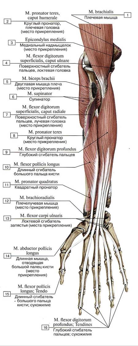
Fig. 321. Third Layer of the anterior muscle group of the right forearm, anterior view:
1 — Brachialis; 2— Pronator teres, humeral head; 3 — Medial epicondyle; 4 — Flexor digitorum superficialis, ulnar head; 5 — Biceps brachii; 6 — Supinator; 7— Flexor digitorum superficialis, radial head; 8 — Pronator teres; 9 — Flexor digitorum profundus; 10 — Flexor pollicis longus; 11 — Pronator quadratus; 12 — Brachioradialis; 13— Flexor carpi ulnaris; 14 — Abductor pollicis longus; 15 — Flexor pollicis longus; Tendon; 16 — Flexor digitorum profundus; Tendons
Function: the flexor digitorum superficialis flexes the middle phalanges of digits II—V, and flexes the hand and forearm.
Blood supply: radial and ulnar arteries.
Innervation: median nerve (CV—ThI).
The third layer of forearm muscles (Figs. 321, 322, 323). The flexor digitorum profundus (m. flexor digitorum profundus) is a broad, flat, triangular muscle. It originates from the proximal two-thirds of the anterior surface of the ulna, inferior to its tuberosity, and from the interosseous membrane of the forearm. The short belly continues into four slender tendons that pass through the carpal tunnel together with the tendons of the flexor digitorum superficialis, subsequently running through the osteofibrous digital sheaths to insert into the bases of the distal phalanges of digits II—V. At the level of the proximal phalanges, each tendon of the flexor digitorum profundus passes through the cleft between the slips of the corresponding superficial flexor tendon (see Fig. 332). This crossing of tendons is known as Camper's chiasm (Petrus Camper, 1722–1789, Dutch anatomist, physician, anthropologist, paleontologist, and artist).
Accessory muscle bundles of the deep flexor of the fingers (Gantzer's accessory heads) arise from the coronoid process of the ulna (Carol Friedrich Ludovic Gantzer, German anatomist. Biographical data are lacking).
The tendons of the finger flexors are connected to the phalanges by fibrous bands—short ligaments of the hand—Marshall's ligaments (John Marshall, 1818–1891, English surgeon and anatomist).
Function: the flexor digitorum profundus flexes the distal phalanges of digits II—V, as well as the hand at the radiocarpal joint.
Blood supply: radial and ulnar arteries.
Innervation: ulnar and median nerves (CV—ThI).
Flexor pollicis longus muscle (m. flexor pollicis longus) is a narrow, fusiform, unipennate muscle located laterally to the deep digital flexor. It originates from the upper two-thirds of the anterior surface of the radius, the adjacent areas of the interosseous membrane of the forearm—extending from the level of the radial tuberosity to the upper border of the pronator quadratus—and from the medial epicondyle of the humerus. The muscle continues into a long tendon that passes through the carpal tunnel within a separate synovial sheath, and then along the palmar side of the thumb between the superficial and deep heads of the flexor pollicis brevis. The tendon inserts into the base of the distal phalanx of the thumb (see Figs. 321, 322, 323).

Fig. 322. Anterior muscle group of the right forearm (transverse section at the middle third of the forearm), anterior view:
1 — Abductor pollicis longus; 2 — Flexor pollicis longus; 3 — Radius; 4 — Extensor carpi radialis brevis; 5 — Extensor carpi radialis longus: 6 — Brachioradialis; 7 — Biceps brachii; Tendon; 8 — Brachialis; 9 — Biceps brachii; 10 - Triceps brachii; 11 — Medial epicondyle, caput commune of flexors; 12 — Bicipital aponeurosis; 13— Pronator teres; 14— Flexor carpi radialis; 15— Palmaris longus; 16— Ulna; 17— Flexor carpi ulnaris; 18 — Flexor digitorum superficialis; 19 — Palmaris longus; Tendon; 20 — Flexor retinaculum

Fig. 323. Anterior muscle group of the right forearm, third layer (A — deep digital flexor, anterior view, superficial forearm muscles removed; B — origin and insertion sites of the anterior forearm muscle group — superficial flexors and pronator teres (diagram)):
1 — Adductor pollicis; 2 — Flexor pollicis longus; Tendon; 3 — Flexor pollicis brevis; 4 — Opponens pollicis; 5 — Pronator quadratus; 6 — Flexor pollicis longus; 7— Pronator teres; 8— Extensor carpi radialis longus; 9 — Brachioradialis; 10— Supinator; 11— Brachialis; 12— Medial epicondyle; 13 — Flexor digitorum profundus; 14 - Flexor carpi ulnaris; 15 — Pisiform; 16 — Opponens digiti minimi; 17 — Flexor digitorum
profundus; Tendons; 18 —- Flexor digitorum superficialis; Tendons
Function: the flexor pollicis longus flexes the distal phalanx of the thumb and the thumb itself, and assists in wrist flexion.
Blood supply: radial, ulnar, and anterior interosseous arteries.
Innervation: median nerve (CV—ThI).
Fourth (deep) layer of forearm muscles (Fig. 324). The pronator quadratus muscle (m. pronator quadratus) is a flat, quadrilateral sheet with transversely oriented muscle fiber bundles. It lies deep to the tendons of the digital and wrist flexors on the anterior surface of the distal third of the forearm bones and the corresponding part of the interosseous membrane of the forearm. The muscle originates from the anterior border and anterior surface of the distal third of the shaft of the ulna, and inserts into the anterior surface of the distal third of the shaft of the radius.
Function: the pronator quadratus rotates the radius around the ulna (pronates the forearm and hand).
Blood supply: anterior interosseous artery.
Innervation: median nerve (CV—ThI).
Posterior muscle group of the forearm. The muscles of the posterior forearm group form two layers—superficial and deep. The superficial layer contains the extensor carpi radialis longus, extensor carpi radialis brevis, extensor digitorum, extensor digiti minimi, and extensor carpi ulnaris. The deep layer comprises the supinator, abductor pollicis longus, extensor pollicis brevis, extensor pollicis longus, and extensor indicis. Each muscle is surrounded by its own fascia.
Superficial layer of the posterior forearm muscle group (Figs. 325, 326).
Extensor carpi radialis longus muscle (m. extensor carpi radialis longus) is an elongated, flat muscle originating with muscular bundles from the lateral epicondyle of the humerus and the lateral intermuscular septum of the arm. At the middle of the forearm, the muscle transitions into a flat tendon that passes into the hand through the second osteofibrous canal (from the lateral side) beneath the extensor retinaculum (together with the extensor carpi radialis brevis). The tendon inserts into the dorsal surface of the base of the second metacarpal bone. The proximal part of the muscle lies adjacent to the lateral surface of the elbow Joint Capsule, after which the muscle extends vertically downwards between the brachioradialis (anteriorly) and the extensor carpi radialis brevis (posteriorly).

Fig. 324. Pronator quadratus and pronator teres, right forearm, anterior view (other anterior forearm muscles removed):
1 — Medial epicondyle; 2 — Joint capsule; Articular capsule; 3 — Biceps brachii; Tendon; 4 — Pronator teres; 5 — Supinator; 6 — Ulna; 7 — Radius; 8 — Interosseous membrane of forearm; 9 — Pronator quadratus

Fig. 325. Posterior muscle group of the right forearm, superficial layer, posterior view:
1 — Extensor digitorum; Tendons; 2 — Intertendinous connections; 3 — Extensor digiti minimi; 4 — Flexor carpi ulnaris; 5 — Anconeus; 6 — Olecranon; 7 — Triceps brachii; 8 — Brachioradialis; 9 — Extensor carpi radialis brevis; 10 — Extensor carpi radialis longus; 11 — Extensor digitorum; 12 — Extensor carpi ulnaris; 13 — Abductor pollicis longus; 14 — Extensor pollicis brevis; 15 — Radius, dorsal tubercle; 16 — Extensor pollicis longus; Tendon

Fig. 326. Brachioradialis, long and short radial wrist extensors, lateral view (A — general view, B — sites of origin and insertion (diagram)):
1 - Metacarpal [III]; 2 — Base of metacarpal [III]; 3 — Radius; 4 — Ulna; 5 — Olecranon; 6 — Lateral epicondyle; 7 — Lateral supraepicondylar ridge; Lateral supracondylar ridge; 8 — Humerus; 9 — Brachioradialis; 10 — Extensor carpi radialis longus; 11 — Extensor carpi radialis brevis; 12 — Radial styloid process; 13 — Base of metacarpal [II]; 14 - Metacarpal [II]
Function: the extensor carpi radialis longus extends the hand and, contracting simultaneously with the flexor carpi radialis, abducts the hand laterally and slightly flexes the forearm.
Blood supply: radial artery, radial collateral and recurrent radial arteries.
Innervation: radial nerve (CV—CVIII).
The extensor carpi radialis brevis (m. extensor carpi radialis brevis) is a fusiform muscle located adjacent (medially) to the extensor carpi radialis longus on the radial side of the forearm. It originates from the lateral epicondyle of the humerus, the radial collateral ligament, and the forearm fascia. At the middle of the forearm, the muscle belly transitions into a long, flat tendon that inserts onto the dorsal surface of the base of the third metacarpal bone. In the distal forearm, the tendons of the short and long wrist extensors pass deep to the more superficially lying, obliquely oriented tendons of the abductor pollicis longus and extensor pollicis longus muscles. In the region of the wrist joint, the tendons of the long and short radial wrist extensors pass within a common (second) osteofibrous canal, enclosed in the synovial Sheath of the radial wrist extensors.
Function: the extensor carpi radialis brevis extends the hand. Contracting simultaneously with the flexor carpi radialis, this muscle abducts the hand.
Blood supply: collateral and recurrent radial arteries.
Innervation: radial nerve (CV—CVIII).
The extensor digitorum (m. extensor digitorum) is a broad, flat muscle positioned superficially, medial to the long and short radial wrist extensors (Fig. 327, see Fig. 325). The extensor digitorum originates from the lateral epicondyle of the humerus and the forearm fascia. Near the wrist joint, this muscle divides into four tendons that pass beneath the extensor retinaculum within a common fourth osteofibrous and synovial sheath (together with the tendon of the extensor indicis). The tendons of the extensor digitorum insert onto the dorsal aspect of digits II–V, forming dorsal digital expansions (extensor hoods). The middle bundles of the expansion insert onto the base of the middle phalanx, while the lateral bundles insert onto the distal (terminal) phalanx. At the level of the metacarpal bones, the tendons of the extensor digitorum are interconnected by obliquely oriented fibrous bands known as intertendinous connections (connexus intertendinei).
A structural variant of the forearm muscles, known as the extensor brevis digitorum manus (or Pozzi's muscle), consists of muscle fibers blending in the lower third of the forearm with the tendons of the extensor digitorum (Samuel Jean Pozzi, 1846–1918, a French physician and anatomist).
Function: the extensor digitorum extends digits II–V and the hand at the wrist joint.
Blood supply: posterior interosseous artery.
Innervation: radial nerve (CV—CVIII).
The extensor digiti minimi (m. extensor digiti minimi) is a narrow, slender muscle that originates from the lateral epicondyle of the humerus and the forearm fascia (together with the extensor digitorum) and lies medial to it. Its narrow, slender tendon passes beneath the extensor retinaculum through a separate fifth osteofibrous and synovial sheath. The tendon inserts onto the dorsal surface of the base of the middle and distal phalanges of the little finger (digit V), blending into their dorsal digital expansions (see Fig. 327).
Function: the muscle extends the little finger.
Blood supply: posterior interosseous artery.
Innervation: radial nerve (CV—CVIII).
The extensor carpi ulnaris (m. extensor carpi ulnaris) is a slender, flat muscle located on the medial side of the dorsal aspect of the forearm, between the extensor digiti minimi (laterally) and the Water/144.html">Origin of the flexor carpi ulnaris (medially). The extensor carpi ulnaris originates from the lateral epicondyle of the humerus, the posterior surface of the ulna, the capsule of the elbow joint, and the forearm fascia (see Fig. 327). The muscle tendon passes through a separate sixth osteofibrous canal on the dorsum of the hand and within its synovial sheath beneath the extensor retinaculum, running in a groove located on the posterior surface of the distal end of the ulna. The extensor carpi ulnaris inserts onto the posterior surface of the base of the fifth metacarpal bone. A bursa is located between THE ORIGIN OF the muscle (its proximal tendon) and the lateral epicondyle of the humerus.
Function: the extensor carpi ulnaris extends and adducts the hand.
Blood supply: posterior interosseous artery.
Innervation: radial nerve (CVI—CVIII).
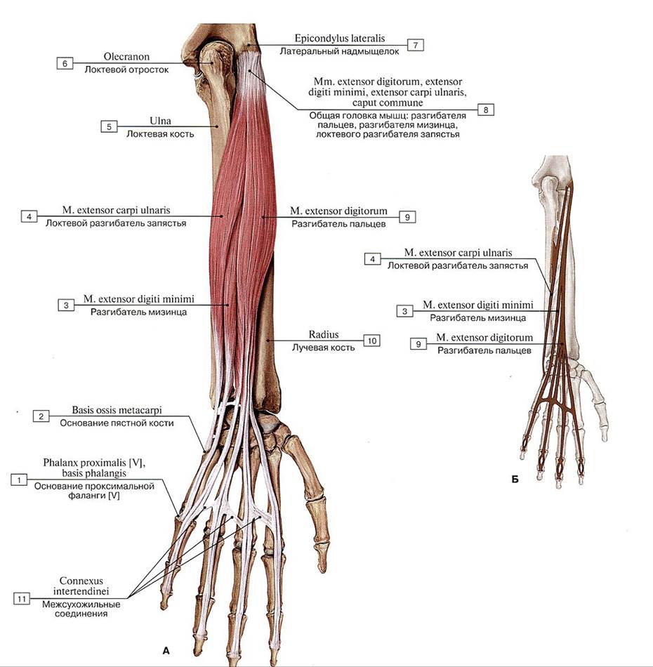
Fig. 327. Extensor muscles of the fingers and extensor carpi ulnaris (A — general view, B — sites of origin and insertion (diagram)):
1 — Proximal phalanx [V], base of phalanx; 2 — Base of metacarpal; 3 — Extensor digiti minimi; 4 — Extensor carpi ulnaris; 5 — Ulna; 6 — Olecranon; 7 — Lateral epicondyle; 8 — Extensor digitorum, Extensor digiti minimi, Extensor carpi ulnaris; Common head; 9 — Extensor digitorum; 10 — Radius; 11— lntertendinous connections
Deep Layer of the forearm muscles (Figs. 328, 329). The supinator muscle (m. supinator) is a flat muscle located in the proximal aspect of the dorsal forearm, almost completely covered by the superficial muscles. The supinator originates from the lateral epicondyle of the humerus, the radial collateral ligament, the annular ligament of the radius, and the supinator crest of the ulna. It passes obliquely in a lateral direction, wrapping around the radius posteriorly and laterally. The muscle inserts into the anterolateral surface of the upper third of the radius, extending from its tuberosity to the insertion site of the pronator teres. The terminal portion of this muscle (anterolateral) is covered by muscles originating from the lateral epicondyle of the humerus (brachioradialis, extensor carpi radialis longus, etc.) and the distal part of the pronator teres. The deep branch of the radial nerve passes longitudinally through the supinator. This slit between the deep and superficial PARTS OF THE supinator is known as the supinator canal (canalis supinatorius).
Function: the supinator rotates the radius outwardly (supinates the radius along with the hand).
Blood supply: radial artery, radial recurrent and interosseous recurrent arteries.
Innervation: radial nerve (CV—CVlll).
The abductor pollicis longus muscle (m. abductor pollicis longus) is a flattened, bipennate muscle. It is located in the distal half of the dorsal forearm between the extensor carpi ulnaris and the supinator, resting on the interosseous membrane and the posterior aspect of the radius. The origin of the abductor pollicis longus is covered posteriorly by the extensor digitorum and the extensor carpi radialis brevis. It originates from the posterior surfaces of the ulna and radius, and the interosseous membrane of the forearm. The long tendon of this muscle runs together with the tendon of the abductor pollicis brevis (correctly: extensor pollicis brevis) in a common first osteofibrous canal on the dorsum of the hand, enclosed in its synovial sheath beneath the lateral part of the extensor retinaculum. The tendon inserts into the dorsal surface of the base of the first metacarpal bone. Distally, the muscle lies directly beneath the forearm fascia and Skin. It courses downward and laterally, curving around the exterior of the radius and the overlying tendons of the long and short radial wrist extensors.
Function: abducts the thumb and the hand.
Blood supply: radial artery, posterior interosseous artery.
Innervation: radial nerve (CV—CVIII).
The extensor pollicis brevis muscle (m. extensor pollicis brevis) is a slender, fusiform muscle situated adjacent (medially and inferiorly) to the abductor pollicis longus. It originates from the posterior surface of the radius and the interosseous membrane. The muscle courses obliquely in a lateral direction across the radius and the overlying tendons. Its long, slender tendon passes beneath the extensor retinaculum within the same (first) synovial sheath as the tendon of the abductor pollicis longus. The tendon inserts into the base of the proximal phalanx of the thumb.
Function: extends the proximal phalanx of the thumb (as well as the digit) and abducts the thumb.
Blood supply: radial artery, posterior interosseous artery.
Innervation: radial nerve (CV—CVI|I).
The extensor pollicis longus muscle (m. extensor pollicis longus) is a slender, fusiform muscle located deep to the extensor digitorum. It originates from the lateral side of the posterior surface of the middle third of the ulna and the interosseous membrane of the forearm. The tendon of this muscle passes beneath the extensor retinaculum through the third osteofibrous canal and its synovial sheath, lying within a groove on the posterior surface of the radius. The tendon of the extensor pollicis longus crosses obliquely and laterally over the tendons of the extensor carpi radialis longus and brevis, and inserts into the base of the distal phalanx of the thumb.
Function: extends the thumb and the hand.
Blood supply: radial artery, posterior interosseous artery.
Innervation: radial nerve (CV—CVIII).
The extensor indicis muscle (m. extensor indicis) is a slender, long, bipennate muscle located on the ulnar side of the forearm adjacent (medially) to the extensor pollicis longus (see Fig. 329). It originates from the posterior surface of the ulna and the interosseous membrane of the forearm. The tendon of this muscle, together with the tendon of the extensor digitorum, travels through the common (fourth) osteofibrous canal and synovial sheath beneath the extensor retinaculum. The tendon inserts into the dorsal surface of the proximal phalanx of the index finger, where it blends with the tendon of the extensor digitorum of that digit.
Function: extends the index finger and assists in extension of the hand.
Blood supply: anterior and posterior interosseous arteries.
Innervation: radial nerve (CV—CVIII).
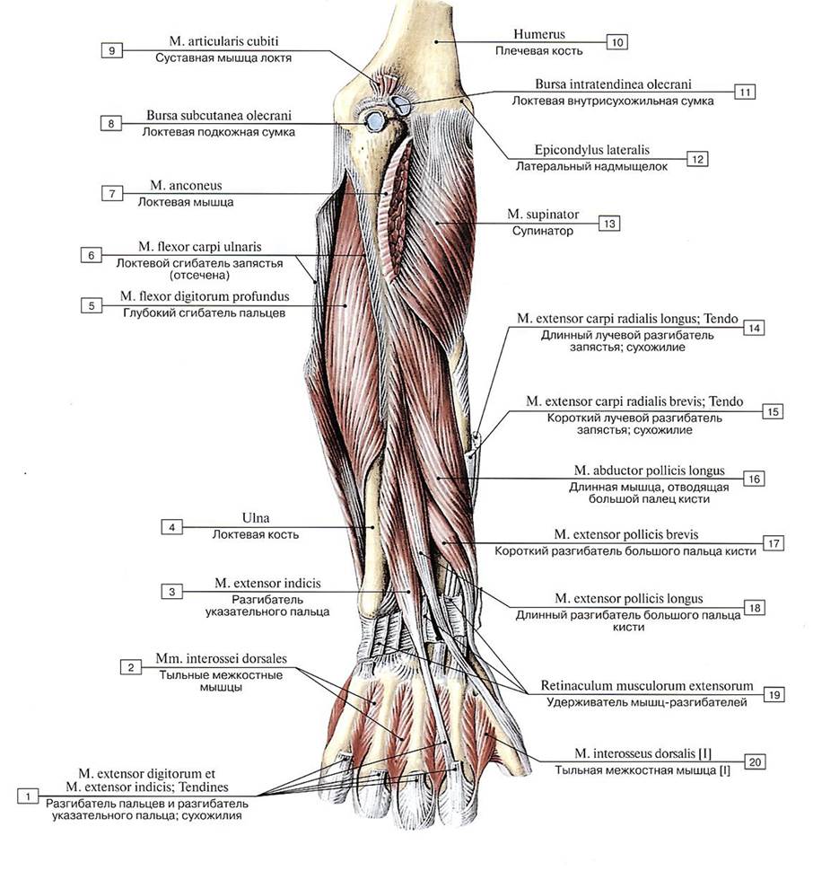
Fig. 328. Posterior muscle group of the right forearm and hand, deep layer, posterior view:
1 — Extensor digitorum and Extensor indicis; Tendons; 2 — Dorsal interossei; 3 — Extensor indicis; 4 — Ulna; 5 — Flexor digitorum profundus; 6 — Flexor carpi ulnaris; 7 —Anconeus; 8 — Subcutaneous olecranon bursa; 9 — Articularis cubiti; 10 — Humerus; 11 — Intratendinous olecranon bursa; 12 — Lateral epicondyle; 13 — Supinator; 14 — Extensor carpi radialis longus; Tendon; 15 — Extensor carpi radialis brevis; Tendon; 16 — Abductor pollicis longus; 17—Extensor pollicis brevis; 18 — Extensor pollicis longus; 19 — Extensor retinaculum; 20 — Dorsal interosseus [I]

Fig. 329. Posterior muscle group of the right forearm, deep layer:
1 — Extensor digiti minimi; 2 — Extensor carpi radialis brevis; Tendon; 3 — Extensor carpi ulnaris; 4 — Flexor carpi ulnaris; 5 — Flexor digitorum profundus; 6 — Anconeus; 7— Medial epicondyle; Common flexor tendon; 8— Triceps brachii; 9 — Brachioradialis; 10 — Extensor carpi radialis longus; 11 - Extensor carpi radialis brevis; 12 — Supinator; 13 — Abductor pollicis longus; 14 — Extensor pollicis longus; 15— Extensor pollicis brevis; 16—Extensor indicis; 17— Extensor carpi radialis longus; Tendon; 18— Extensor digitorum
Last update: 08/08/2026
Editorial and Educational Adaptation: This material has been compiled based on the primary/original source text. The project team performed an editorial review, corrected technical inaccuracies, structured sections, and adapted the content for an educational format.
What was processed:
- elimination of formatting defects (OCR errors, structural breaks, corrupted characters);
- editorial organization of content;
- standardization of terminology in accordance with academic sources;
- verification of factual statements against the original source text.
All mentions of the author, publication year, and origin of the primary text have been preserved in accordance with the source.