HUMAN ANATOMY ATLAS - G. L. Bilich - 2014
Muscular System
MUSCLES OF THE SHOULDER GIRDLE
The Muscles of the shoulder girdle (Pectoral Girdle) surround the shoulder joint anteriorly, superiorly, and posteriorly. The superficial layer is formed by the deltoid Muscle, while the deep layer comprises the supraspinatus, infraspinatus, teres major, teres minor, and subscapularis muscles.
The deltoid muscle (m. deltoideus) is a thick, convex, triangular plate located directly beneath the Skin. This muscle covers the shoulder joint from the front, top, back, and lateral side, creating the rounded contour of the shoulder. The multipennate deltoid muscle originates by a short tendon from the anterior border of the lateral third of the clavicle, the lateral margin of the acromion, the spine of the scapula, and, partially, from the infraspinatous fascia. The fascicles of its three portions (corresponding to their origin), separated from one another by Connective Tissue septa, converge laterally toward the lateral aspect of the humerus and insert into its deltoid tuberosity (see Figs. 303, 304, 305, 306). The superficial fascicles of the deltoid muscle are fleshy, whereas the deep ones are tendinous. The anterior border of the deltoid muscle borders the superior margin of the pectoralis major muscle. Between these muscles lies a narrow, triangular deltopectoral groove (sulcus deltoideopectoralis), which widens near the clavicle. The subdeltoid bursa (bursa subdeltoidea) is situated beneath the deltoid muscle, between the deep lamina of the Fascia of the same name and the greater tubercle of the humerus; in approximately 10% of cases, it communicates with the cavity of the shoulder joint.
As an anatomical variant, the scapulocostodeltoid muscle—known as Calori's muscle—arises from the lateral border of the scapula and inserts into the deltoid muscle or its aponeurosis. This muscle is highly variable in shape and attachment (Luigi Calori, 1807–1896, an Italian anatomist).
Function: The deltoid muscle abducts the arm from the trunk up to the horizontal level; its anterior clavicular part flexes the shoulder, medially rotating it, and lowers the raised arm; its posterior part extends the shoulder, laterally rotating it simultaneously, and lowers the raised arm.
Innervation: Axillary nerve (CV—CVI).
Blood supply: Thoracoacromial artery, posterior circumflex humeral artery.
The supraspinatus muscle (m. supraspinatus) is a flat, triangular plate located in the supraspinous fossa and covered superiorly by the trapezius muscle. It originates from The surface of the supraspinous fossa of the scapula and the supraspinatous fascia. The muscle fascicles pass laterally and insert by a short tendon into the superior facet of the greater tubercle of the humerus (Figs. 307, 308, 309, 310). The tendon passes beneath the coracoid process and the coracohumeral ligament, where it blends with the capsule of the shoulder joint.
Function: The supraspinatus muscle abducts the shoulder when the scapula is fixed, acting as a synergist to the deltoid muscle; it pulls the capsule of the shoulder joint upward, protecting it from pinching.
Blood supply: Suprascapular artery, circumflex scapular artery.
Innervation: Suprascapular nerve (CV—CVI).
The infraspinatus muscle (m. infraspinatus) is a flat, triangular plate located in the infraspinous fossa, covered by the deltoid and trapezius muscles. It originates from the surface of the infraspinous fossa of the scapula and the infraspinatous fascia. The muscle fascicles converge laterally and superiorly, pass posterior to the shoulder joint, and insert into the middle posterior facet of the greater tubercle of the humerus via a short tendon (see Figs. 307, 309, 310). In the area of attachment to the bone, There is a subtendinous infraspinatus synovial bursa (bursa subtendinea musculi infraspinati).
Function: The infraspinatus muscle laterally rotates (supinates) the shoulder and adducts it toward the trunk, and pulls on the Joint Capsule, into which some of its fascicles blend.
Class="center">
Fig. 303. Muscles of the right upper limb (fasciae removed), anterior view:
1 — Flexor digitorum profundus; Tendons; 2 — - Tendon sheath; 3 — Thenar eminence; 4 — Extensor carpi radialis longus; 5 — Brachioradialis; 6 — Deltoid; 7 — Clavicle; 8 — Pectoralis major; 9 — Biceps brachii; 10 — Braehialis; 11 — Pronator teres; 12— Flexor carpi radialis; 13 — Palmaris longus; Tendon; 14 — Flexor digitorum superficial is; Tendon; 15 — Flexor carpi ulnaris; Tendon; 16 — Flexor retinaculum; 17— Hypothenar eminence; 18 — Flexor digitorum superficialis; Tendons

Fig. 304. Muscles of the right upper limb and chest, lateral view:
1— Dorsal interossei; 2 — Extensor pollicis longus: Tendon; 3 — Extensor retinaculum; 4 — Extensor pollicis brevis; 5 — Extensor digitorum; Tendon; 6 — Radius; 7— Extensor carpi radialis brevis; 8— Extensor digitorum; 9 — Anconeus; 10 — Olecranon; 11 — Lateral intermuscular septum of arm; 12 — Triceps brachii; Tendon; 13 — Latissinius dorsi; 14 — Triceps brachii, lateral HEAD; 15 — Triceps brachii, long head; 16 — Teres major; 17— Teres minor; 18— Infraspinous fascia; 19 — Trapezius; 20— Spine of scapula; 21 — Acromion; 22— Sternocleidomastoid; 23 — Clavicle; 24 — Pectoralis major; 25 — Deltoid; 26 — Pectoralis major, abdominal part; 27 — Serratus anterior; 28 — Rectus sheath, anterior layer; 29 —External oblique; 30 — Biceps brachii; 31 — Brachialis; 32— Lateral epicondyle; 33— Brachioradiaiis; 34 — Extensor carpi radialis longus; 35 — Flexor carpi radialis; 36 — Flexor pollicis longus; 37 — Abductor pollicis longus; 38— Abductor pollicis longus; Tendon; 39— Extensor pollicis brevis; Tendon; 40 — Adductor pollicis

Fig. 305. Muscles of the right upper limb (fasciae and extensor retinaculum removed), posterior view:
1— Extensor carpi ulnaris; 2 — Anconeus; 3 — Teres major; 4 — Teres minor; 5 — Infraspinatus; 6 — Trapezius; 7— Acromion; 8 — Deltoid; 9 — Triceps brachii; 10 — Brachioradialis; 11— Extensor carpi radialis longus; 12— Extensor carpi radialisbrevis; 13— Extensor digitorum; 14 — Dorsal interossei
Blood supply: Circumflex scapular artery, suprascapular artery.
Innervation: suprascapular nerve (CV—CVI).
The teres minor muscle is an elongated, cylindrical muscle positioned inferior to the infraspinatus and partially covered by the scapular part of the deltoid muscle (Fig. 310, see Fig. 307). It originates from the lateral border of the scapula and the infraspinous fascia, courses superiorly and laterally, and inserts into the lower posterior facet of the greater tubercle of the humerus (see Fig. 309). Some of its muscle bundles blend into the capsule of the shoulder joint.
Action: The teres minor laterally rotates (supinates) the arm, acting as a synergist to the subscapularis and the scapular part of the deltoid; it also pulls on the capsule of the shoulder joint, preventing its impingement.
Blood supply: circumflex scapular artery and suprascapular artery.
Innervation: suprascapular nerve (CV).
Table 46. Muscles of the Upper Limb
Muscle name |
Origin |
Insertion |
Action |
Innervation, blood supply |
|
Muscles of the shoulder girdle |
|||||
Deltoid muscle |
Acromial end of clavicle, acromion, spine of scapula |
Deltoid tuberosity of humerus |
Entire muscle abducts the arm; anterior part flexes the arm; posterior part extends the arm |
Axillary nerve (CV-CVI). Aa.: posterior circumflex humeral; thoracoacromial |
|
Supraspinatus muscle |
Supraspinous fossa of scapula, supraspinous fascia |
Greater tubercle of humerus, capsule of shoulder joint |
Abducts the arm, pulls on the capsule of the shoulder joint |
Suprascapular nerve (CV-CVl). Aa.: suprascapular; circumflex scapular |
|
Infraspinatus muscle |
Infraspinous fossa, infraspinous fascia |
Greater tubercle of humerus |
Laterally rotates the arm |
||
Teres minor muscle |
Lateral border of scapula, infraspinous fascia |
Greater tubercle of humerus |
Laterally rotates the arm |
Axillary nerve (CV-CVI). Aa.: circumflex scapular |
|
Teres major muscle |
Inferior angle of scapula, infraspinous fascia |
Crest of lesser tubercle of humerus |
Extends the arm, medially rotates it |
Subscapular nerve (CV-CVl). Aa.: subscapular |
|
Subscapularis muscle |
Costal surface of scapula |
Lesser tubercle of humerus |
Medially rotates the arm and adducts it to the trunk |
||
Muscles of the Free Upper Limb Muscles of the arm Anterior Group of arm muscles |
|||||
Coracobrachialis muscle |
Coracoid process of scapula |
Humerus inferior to the crest of the lesser tubercle |
Flexes the arm at the shoulder joint and adducts it |
Musculocutaneous nerve (СV-СVIII). Aa.: anterior and posterior circumflex humeral |
|
Biceps brachii |
Supraglenoid tubercle of scapula (long head), coracoid process of scapula (short head) |
Tuberosity of radius |
Flexes and supinates the forearm at the elbow joint, flexes the arm at the shoulder joint |
Musculocutaneous nerve (СV-СVIII). Aa.: brachial; superior and inferior ulnar collateral; radial recurrent |
|
Brachialis muscle |
Humerus, distal to the deltoid tuberosity |
Same |
Flexes the forearm at the elbow joint |
||
Posterior group of arm muscles |
|||||
Triceps brachii |
Infraglenoid tubercle of scapula (long head), posterior surface of humeral shaft (medial and lateral heads) |
Olecranon of ulna |
Extends the forearm at the elbow joint. The long head extends and adducts the arm at the shoulder joint |
Radial nerve (СV-СVIII). Aa.: superior and inferior ulnar collateral; posterior circumflex humeral; profunda brachii a. |
|
Anconeus muscle |
Lateral epicondyle of humerus |
Olecranon, posterior surface of ulna |
Extends the forearm at the elbow joint |
Radial nerve (СV-СVIII). Aa.: recurrent interosseous |
|
Muscles of the forearm Anterior group of forearm muscles Superficial layer of forearm muscles |
|||||
Brachioradialis muscle |
Lateral supracondylar ridge of humerus, lateral intermuscular septum of arm |
Radius superior to the styloid process |
Flexes the forearm, placing it in a mid-position between pronation and supination |
Radial nerve (СV-СVIII). Aa.: radial; radial collateral; radial recurrent |
|
Pronator teres |
Medial epicondyle of humerus, coronoid process of ulna |
Lateral surface of radius |
Pronates and flexes the forearm |
Median nerve (CV-ThI). Aa.: brachial; ulnar; radial |
|
Flexor carpi radialis |
Medial epicondyle of humerus, medial intermuscular septum of arm, forearm fascia |
Palmar surface of base of Metacarpal bones I–II |
Flexes the wrist and abducts the hand, flexes the forearm |
Median nerve (CV-ThI). Aa.: radial; ulnar; brachial |
|
Palmaris longus |
Medial epicondyle of humerus, medial intermuscular septum of arm |
Palmar aponeurosis |
Tenses the palmar aponeurosis, flexes the hand and forearm |
Median nerve (CV-ThI). Aa.: radial |
|
Flexor carpi ulnaris |
Medial epicondyle of humerus, medial intermuscular septum of arm, olecranon of ulna, forearm fascia |
Pisiform and hamate bones, base of metacarpal bone V |
Flexes the wrist and adducts the hand, flexes the forearm |
Ulnar nerve (CVII-ThI). Aa.: ulnar; superior and inferior ulnar collateral |
|
Second layer of forearm muscles |
|||||
Flexor digitorum superficialis |
Medial epicondyle of humerus, coronoid process of ulna, anterior border of radius, forearm fascia |
Four tendons attach to the palmar surface of the middle Phalanges of digits II–V |
Flexes the middle phalanges of digits II–V, flexes the wrist and forearm |
Median nerve (CV-ThI). Aa.: radial; ulnar |
|
Third Layer of forearm muscles |
|||||
Flexor digitorum profundus |
Anterior surface of ulna, interosseous membrane of forearm |
Four tendons attach to the distal phalanges of the digits |
Flexes the distal phalanges of digits II–V, flexes the wrist |
Median nerve (CVII-ThI). Aa.: radial; ulnar |
|
Flexor pollicis longus |
Anterior surface of radius, interosseous membrane of forearm |
Palmar surface of distal phalanx of thumb (digit 1) |
Flexes the thumb, flexes the wrist |
Median nerve (CV-ThI). Aa.: radial; anterior interosseous |
|
Fourth layer of forearm muscles |
|||||
Pronator quadratus |
Anterior border and anterior surface of ulna |
Anterior surface of radius |
Pronates the forearm and hand |
Median nerve (CV-ThI). Aa.: anterior interosseous |
|
Posterior group of forearm muscles Superficial layer of forearm muscles |
|||||
Extensor carpi radialis longus |
Lateral epicondyle of humerus, lateral intermuscular septum of arm |
Dorsal surface of base of metacarpal bone II |
Extends the wrist, abducts it radially, flexes the forearm |
Radial nerve (CV-CVIII). Aa.: radial; radial collateral; radial recurrent |
|
Extensor carpi radialis brevis |
Lateral epicondyle of humerus, forearm fascia |
Dorsal surface of base of metacarpal bone III |
Extends and abducts the wrist |
||
Extensor digitorum |
Lateral epicondyle of humerus, forearm fascia |
Dorsal surface of middle and distal phalanges of digits II–V |
Extends digits II–V, extends the wrist |
Radial nerve (CV-CVIII). Aa.: posterior interosseous |
|
Extensor digiti minimi |
Lateral epicondyle of humerus, forearm fascia |
Dorsal surface of middle and distal phalanges of the little finger |
Extends the little finger |
||
Extensor carpi ulnaris |
Lateral epicondyle of humerus, forearm fascia |
Dorsal surface of base of metacarpal bone V |
Extends and adducts the wrist |
Radial nerve (CV-CVIII). Aa.: posterior interosseous |
|
Deep Layer of forearm muscles |
|||||
Supinator |
Lateral epicondyle of humerus, ulna |
Proximal third of lateral surface of radius |
Supinates the forearm |
Radial nerve (CV-CVIII). Aa.: radial; recurrent; recurrent interosseous |
|
Abductor pollicis longus |
Posterior surface of ulna and radius, interosseous membrane of forearm |
Dorsal surface of base of metacarpal bone I |
Abducts the thumb and wrist |
Radial nerve (CV-CVIII). Aa.: radial; posterior interosseous |
|
Extensor pollicis brevis |
Posterior surface of radius, interosseous membrane of forearm |
Dorsal surface of base of proximal phalanx of thumb |
Extends the proximal phalanx of the thumb |
||
Extensor pollicis longus |
Posterior surface of ulna, interosseous membrane of forearm |
Dorsal surface of base of distal phalanx of thumb |
Extends the thumb |
Radial nerve (CV-CVIII). Aa.: radial; posterior interosseous |
|
Extensor indicis |
Posterior surface of ulna, interosseous membrane of forearm |
Dorsal surface of proximal phalanx of index finger |
Extends the index finger |
Radial nerve (CV-CVIII). Aa.: posterior interosseous |
|
|
Thenar muscles |
|||||
Abductor pollicis brevis |
Scaphoid bone, trapezium, flexor retinaculum |
Lateral margin of base of proximal phalanx of thumb |
Abducts the thumb |
Median nerve (CV-ThI). Aa.: superficial palmar branch of radial a. |
|
Flexor pollicis brevis |
Trapezium, trapezoid bone, flexor retinaculum, metacarpal bone II |
Anterior surface of base of proximal phalanx of thumb |
Flexes the thumb |
Superficial head: median nerve (CV-ThI), deep head: ulnar nerve (CVII-ThI). Aa.: superficial palmar branch of radial a.; deep palmar arch |
|
Opponens pollicis |
Trapezium, flexor retinaculum |
Lateral margin and anterior surface of metacarpal bone I |
Opposes the thumb to the little finger |
Median nerve (CV-ThI). Aa.: superficial palmar branch of radial a.; deep palmar arch |
|
Adductor pollicis |
Capitate bone, base and anterior surface of metacarpal bones II and III |
Base of proximal phalanx of thumb |
Adducts the thumb |
Ulnar nerve (CVII-ThI). Aa.: superficial and deep palmar arches |
|
Hypothenar muscles |
|||||
Palmaris brevis |
Flexor retinaculum |
Skin of medial margin of hand |
Wrinkles the skin over the hypothenar eminence |
Ulnar nerve (CVII-ThI). Aa.: ulnar |
|
Abductor digiti minimi |
Flexor retinaculum, pisiform bone |
Medial margin of base of proximal phalanx of little finger |
Abducts the little finger |
Ulnar nerve (CVII-ThI). A.: deep palmar branch of ulnar |
|
Flexor digiti minimi brevis |
Hook of hamate, flexor retinaculum |
Palmar surface of proximal phalanx of little finger |
Flexes the little finger |
Ulnar nerve (CVII-ThI). Aa.: deep palmar branch of ulnar |
|
Opponens digiti minimi |
Flexor retinaculum, hook of hamate |
Medial margin and anterior surface of metacarpal bone V |
Opposes the little finger to the thumb |
Ulnar nerve (CVII-ThI). Aa.: deep palmar branch of ulnar |
|
Central group of muscles |
|||||
Lumbricals |
Tendons of the flexor digitorum profundus |
Dorsal surface of proximal phalanges of digits |
Flex the proximal phalanges, extend the middle and distal phalanges of digits II–V |
I and II muscles: median nerve (CV-ThI), III and IV muscles: ulnar nerve (CVII-ThI). Aa.: superficial and deep palmar arches |
|
Palmar interossei |
Medial side of metacarpal II, lateral side of metacarpals IV and V |
Dorsal surface of proximal phalanges of digits II, IV, and V |
Adduct digits II, IV, and V toward digit III |
Ulnar nerve (CVII-ThI). Aa.: deep palmar arch |
|
Dorsal interossei |
Adjacent sides of metacarpal bones I–V |
Dorsal surface of proximal phalanges of digits II, III, and IV |
Abduct digits II, IV, and V away from digit III |
Ulnar nerve (CVII-ThI). Aa.: deep palmar arch; dorsal metacarpal |
|

Fig. 306. Deltoid muscle, right, all other muscles removed (A — lateral view, B — posterior view, C — origin and insertion sites of the deltoid muscle (diagram)):
1 — Shaft of humerus; Body of humerus; 2 — Scapula; 3 — Deltoid, spinal part; 4 — Deltoid, acromial part; 5 — Spine of scapula; 6 — Acromion; 7 — Clavicle; 8 — Deltoid, clavicular part; 9— Deltoid tuberosity; 10— Scapula, costal surface; 11 — Coracoid process
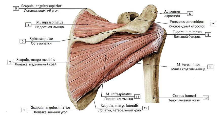
Fig. 307. Muscles of the right shoulder girdle: supraspinatus, infraspinatus, teres minor, subscapularis, posterior view:
1 — Scapula, inferior angle; 2 — Scapula, medial border; Crest of lesser tubercle; Medial lip; 3 — Spine of scapula; 4 — Supraspinatus; 5 - Scapula, superior angle; 6 — Acromion; 7 - Coracoid process; 8 - Greater tubercle; 9 — Teres minor; 10 — Shaft of humerus; Body of humerus; 11 — Infraspinatus; 12— Scapula, lateral border

Fig. 308. Muscles of the right shoulder girdle: supraspinatus, infraspinatus, teres minor, subscapularis, anterior view:
1 - Shaft of humerus; Body of humerus; 2 — Crest of lesser tubercle; Medial lip; 3 — Crest of greater tubercle; Lateral lip; 4 — Coracoid process; 5 —- Intertubercular sulcus; Bicipital groove; 6— Lesser tubercle; 7 — Greater tubercle; 8 — Acromion; 9 — Scapula, superior angle; 10- Supraspinatus; 11 — Scapula, superior border; 12— Suprascapular notch; 13 — Scapula, medial border; 14 — Subscapularis; 15 — Scapula, inferior angle

Fig. 309. Origin and insertion of muscles: supraspinatus, infraspinatus, and teres minor (diagram):
1 — Teres minor; 2 — Infraspinatus; 3 — Supraspinatus
The teres major muscle is cylindrical and somewhat flattened, situated near the lateral border of the scapula and covered by the distal tendon of the latissimus dorsi. It originates from the lower part of the lateral border and the inferior angle of the scapula, as well as from the infraspinous fascia. The muscle inserts via a broad, flat tendon into the crest of the lesser tubercle of the humerus, inferior and slightly posterior to the insertion of the latissimus dorsi tendon (see Fig. 310). The muscle bundles of the teres major course along the lateral border of the scapula, crossing the humerus from the medial side inferior to the level of the surgical neck. A subtendinous bursa of the teres major muscle (bursa subtendinea musculi teretis majoris) is located near the insertion site.
Action: With a fixed scapula, the teres major extends the arm at the shoulder joint, pronates it, and adducts the elevated arm to the trunk. With a fixed upper limb, it pulls the inferior angle of the scapula laterally and displaces it anteriorly.
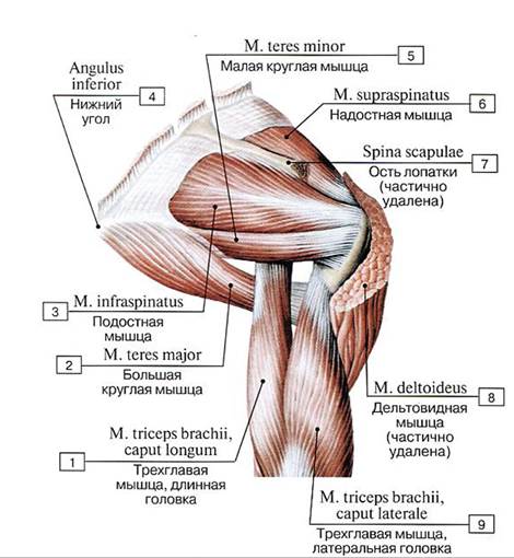
Fig. 310. Muscles of the right upper limb, posterior view:
1 — Triceps brachii, long head; 2 — Teres major; 3 — Infraspinatus; 4 — Inferior angle; 5 — Teres minor; 6 — Supraspinatus; 7— Spine of scapula; 8 — Deltoid; 9 — Triceps brachii, lateral head

Fig. 311. Origin and insertion of the subscapularis muscle (diagram):
1 — Supraspinatus

Fig. 312. Muscles of the left pectoral girdle, posterior view:
1 — Spatium axilare mediale; Triangular space; 2 — Spatium axilare laterale; Quadrangular space; 3 — Triceps brachii, long head; 4 — Triceps brachii, lateral head; 5 — Glenohumeral joint; Shoulder joint; Joint capsule; Articular capsule; 6 — Humerus, surgical neck; 7 — Deltoid; 8 — Subdeltoid bursa; 9 — Acromion; 10 — Omohyoid, inferior belly; 11 — Levator scapulae; 12 — Superior angle; 13 — Rhomboid minor; 14 — Supraspinatus; 15 — Trapezius; 16 — Rhomboid major; 17 — Spine of scapula; 18 — Infraspinatus; 19 — Teres minor; 20 — Teres major; 21 — Inferior angle; 22 — Serratus anterior; 23 — Latissimus dorsi
Innervation: subscapular nerve (CV–CVII).
Blood supply: subscapular artery.
The subscapularis muscle (m. subscapularis) is a thick, triangular muscle located in the subscapular fossa. It arises from the costal surface of the scapula, its lateral margin, and partially from the subscapular fascia via muscular bundles. Passing laterally, the muscle inserts via a flat tendon into the lesser tubercle and the crest of the lesser tubercle of the humerus (Fig. 311, see Fig. 308). The tendon of the subscapularis lies adjacent to the anteroinferior surface of the shoulder joint capsule, and the inferior margin of the muscle contacts the teres major. Between the subscapular tendon and the shoulder joint capsule lies the subtendinous bursa of the subscapularis muscle (bursa subtendinea musculi subscapularis), which typically communicates with the cavity of the shoulder joint.
Function: The subscapularis muscle pronates the arm, medially rotates it, and adducts it toward the trunk.
Blood supply: subscapular artery.
Innervation: subscapular nerve (CV–CVII).
Between the clavicle and the scapula, a small and rare anatomical variant may occasionally be found: Rousseau's muscle (syn.: deep acromioclavicular muscle, m. acromioclavicularis profundus) (named after Louis François Emmanuel Rousseau, 1788–1868, a French anatomist and histologist). Covered by the deltoid muscle, it originates from the clavicle at the junction of its middle and lateral thirds and inserts into the apex of the acromion of the scapula.
Last update: 08/08/2026
Editorial and Educational Adaptation: This material has been compiled based on the primary/original source text. The project team performed an editorial review, corrected technical inaccuracies, structured sections, and adapted the content for an educational format.
What was processed:
- elimination of formatting defects (OCR errors, structural breaks, corrupted characters);
- editorial organization of content;
- standardization of terminology in accordance with academic sources;
- verification of factual statements against the original source text.
All mentions of the author, publication year, and origin of the primary text have been preserved in accordance with the source.