MODERN BOTANY - P. RAVEN - 1990
SECTION III. GENETICS
CHAPTER 9. EUKARYOTIC GENETICS
Meiosis
Meiosis takes place in specialized diploid Cells and only at specific stages of an Organism's life cycle. As a result of meiosis and cytokinesis, a single diploid Cell gives rise to four haploid cells—Gametes or spores. A gamete is a cell that fuses with another gamete to form a diploid zygote. The zygote can then divide either meiotically to form four haploid cells or mitotically to give rise to a multicellular diploid organism. Haploid cells can also function as independent unicellular organisms. If a multicellular diploid organism is formed, it will, in most cases, sooner or later produce haploid spores or gametes via meiosis. A spore is a cell that can develop into a complete organism without fusing with another cell. Spores often divide mitotically, forming a haploid multicellular organism that produces gametes through mitosis (see Fig. 10-11).
First Division of Meiosis
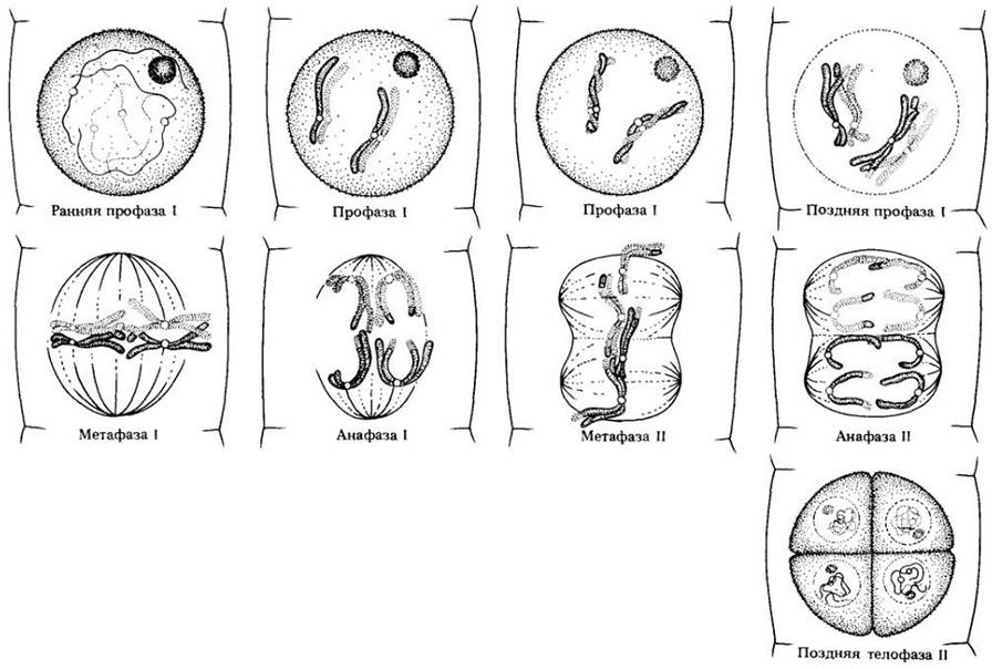
Meiosis consists of two successive nuclear divisions. Fig. 9-6 will help us trace the processes described in the following paragraphs.
In prophase I (the prophase of the first meiotic division), the Chromosomes appear as long, thin threads. As in mitosis (see Chapter 2), the chromosomes have already duplicated during the preceding interphase. Consequently, at the beginning of prophase I, each chromosome consists of two identical chromatids joined by a centromere. However, at this early stage of meiosis, each chromosome appears as a single unified Structure.
Before the chromatids become visible, homologous chromosomes pair up (synapse) with one another. Synapsis occurs very precisely, beginning at one or more points and continuing along the entire length of the chromosome much like a zipper closing. Each homolog is derived from one parent and consists of two identical chromatids. Thus, homologous pairs consist of four chromatids. The pairing of homologous chromosomes is an obligatory part of meiosis; it cannot occur in haploid cells because they lack homologs. The pairing process itself is called synapsis, and the joined pairs of homologous chromosomes are referred to as bivalents.
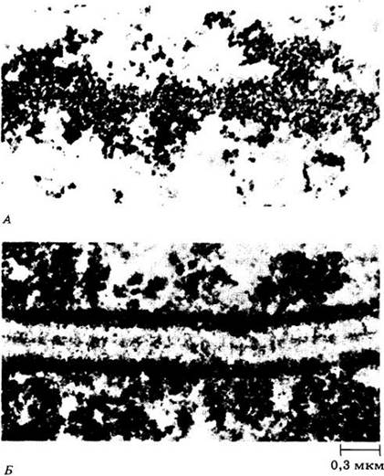
During prophase I, the chromosomes shorten and thicken. Under an Electron microscope, a darkly staining axial protein core can be seen within each chromosome (Fig. 9-3, A). In the middle of prophase, the axial cores of homologous chromosomes approach one another to a final distance of 0.1 µm, forming the synaptonemal complex (Fig. 9-3, B).
Class="center">Fig. 9-3. A. A fragment of a Lilium chromosome in prophase I prior to synapsis. Note the dense axial core, consisting mainly of Proteins. It likely organizes the genetic material, preparing it for pairing and Gene exchange. B. The synaptonemal complex in a Lilium bivalent. Of the four chromatids, only two are visible
In favorable material at this time, it can be observed that each bivalent consists of four chromatids, two in each chromosome. Within the synaptonemal complex, segments are exchanged between homologous chromatids. This process, known as Crossing Over, results in the chromatids acquiring a new gene composition. Fig. 9-4 clearly illustrates The process of crossing over; the visible X-shaped structure is called a chiasma.
Fig. 9-4. Pairing chromosomes of the grasshopper Chorhippus parallelus. Variations in the number of chiasmata can be observed

Toward the end of prophase I, the synaptonemal complex disintegrates. The nuclear envelope then breaks down, and the nucleoli usually disappear as RNA Synthesis halts. The homologous chromosomes separate, but the chromatids remain held together at the chiasmata (points of contact) and pull apart very slowly. As the chromatids separate, some chiasmata shift toward the ends of the chromosome arms. One or more chiasmata may be found in each chromosome arm, or a single chiasma for the entire bivalent; The structure of individual bivalents can vary widely depending on the number of chiasmata (Fig. 9-4).
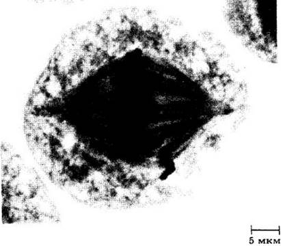
In metaphase I, a spindle-like structure formed by microtubules becomes noticeable (Fig. 9-5). During meiosis, individual microtubules attach to the centromeres of the chromosomes of each bivalent. The chromosome pairs then move to the equatorial plane of The Cell, where they align in random order. The centromeres of homologous chromosomes lie on opposite sides of the equatorial plane; in contrast, during mitotic metaphase, as we have seen, the centromeres of individual chromosomes lie directly on the equatorial plane.
Fig. 9-5. The spindle in a pollen mother cell of wheat (Triticum aestivum) during metaphase I of meiosis
Anaphase I begins with the Separation of homologous chromosomes and their movement toward the poles. (Note again the difference from mitosis: in mitotic anaphase, the centromeres divide and identical chromatids separate.) In meiotic anaphase I, the centromeres do not divide, the chromatids remain together, and the homologous chromosomes separate instead. However, due to the exchange of fragments resulting from crossing over, the chromatids are no longer identical as they were at THE START OF meiosis.
In telophase I, chromosome coiling relaxes, and they lengthen, becoming indistinguishable once again. As telophase gradually transitions into interphase, a nuclear envelope reforms from The Endoplasmic reticulum. Finally, the nucleolus reappears and Protein Synthesis resumes. In many organisms, however, interphase is absent; in this case, the chromosomes transition more or less directly from telophase I to prophase II of the second meiotic division.
Second Division of Meiosis
At the start of the second meiotic division, the chromatids are still connected by their centromeres. This division resembles mitosis: if a nuclear envelope formed in telophase I, it now breaks down, and by the end of prophase II, the nucleolus disappears. In metaphase II, the spindle and chromosomes consisting of two chromatids can be seen once again. The chromosomes are attached by their centromeres to the spindle fibers and line up along the equatorial plane. In anaphase II, the centromeres divide and separate, and the sister chromatids—now individual chromosomes—move toward opposite poles (see Fig. 8-1). In telophase II, new nuclear envelopes and nucleoli form, chromosome Condensation relaxes, and they become invisible within the interphase Nucleus. The overall scheme of meiosis is shown in Fig. 9-6.
Fig. 9-6. Schematic representation of meiosis with two pairs of chromosomes. Not all stages are shown.
Prophase I: chromosomes become visible as long threads; homologous chromosomes approach and pair up, wrapping around each other, and the pairing chromosomes become very short.
Metaphase I: homologous chromosomes align at the equatorial plane, with their centromeres directed toward opposite poles.
Anaphase I: paired chromosomes separate and move toward opposite poles.
The second meiotic division is essentially a mitotic division.
Metaphase II: chromosomes line up along the equatorial plane, with their centromeres positioned on the plate.
Anaphase II: centromeres divide, and chromatids separate and move toward opposite poles.
Telophase II: chromosome migration is complete, and four new nuclei are formed, each with a haploid chromosome number.
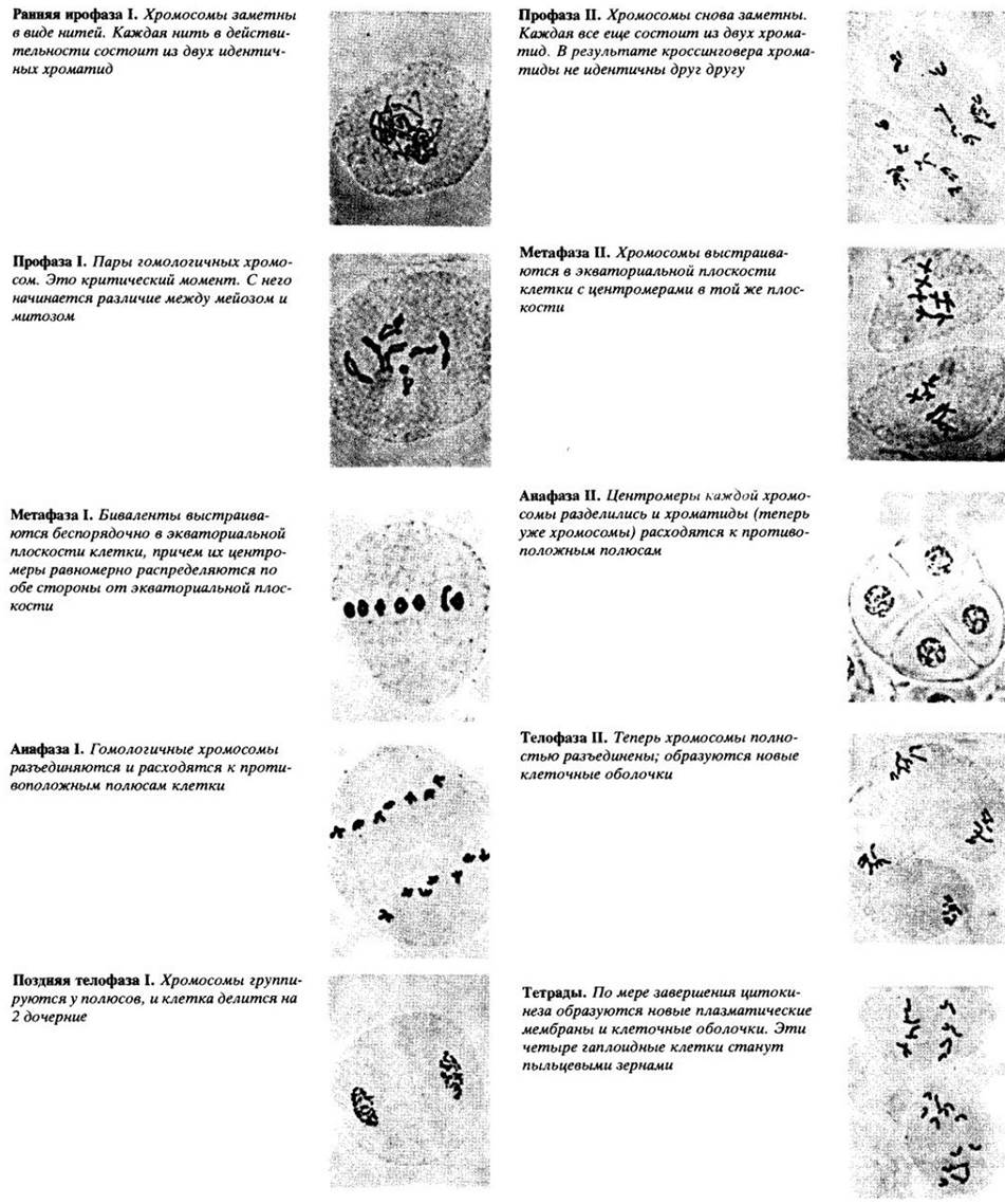
Meiosis in crested wheatgrass (Agropyron cristatum), n = 7, is shown below
Significance of Meiosis
The net result of meiosis is that the genetic material present in a diploid nucleus is replicated only once, but divided twice. Consequently, each daughter cell receives only half the number of chromosomes present in the diploid nucleus. The Genetic consequences of meiosis are even more significant. During metaphase I, the orientation of bivalents is random, meaning that the chromosomes of each parent are distributed at random between the two new nuclei. Furthermore, as a result of crossing over, individual chromosomes frequently consist of chromosomal segments from both parents. If the original diploid cell had two pairs of homologous chromosomes, n = 2, there are four possible ways in which chromosome pairs can align at metaphase, and correspondingly four patterns of their distribution among haploid cells. If n = 3, there are 8 possibilities; if n = 4, 16. The general formula is 2n. In humans, n = 23, so the number of possible combinations is 223, which equals 8,388,608. In many organisms, the chromosome number is even higher than in humans.
As the chromosome number increases, the probability of regenerating a chromosome Complement identical to that of the original diploid nucleus decreases sharply. Moreover, the presence of at least one chiasma in each bivalent makes it virtually impossible for meiosis to produce a cell genetically identical to either of the parental gametes whose fusion originally formed the parent organism. There are three fundamental differences between meiosis and mitosis (see Fig. 9-7):
Fig. 9-7. Comparison of meiosis and mitosis

1. Although the genetic material is duplicated only once during meiosis, two successive divisions occur, leading to The formation of four nuclei.
2. Each of the four resulting nuclei is haploid, containing only half the chromosome number characteristic of the original diploid nucleus.
3. The nuclei produced by meiosis contain novel combinations of chromosomes.
As a result of meiosis, the resulting nuclei differ from the parent nucleus, in contrast to mitosis, where the chromosome sets of the daughter and parent nuclei are identical. The behavior of chromosomes during meiosis has profound genetic and evolutionary implications. Through meiosis and syngamy, natural populations of diploid organisms are far from uniform; instead, they consist of individuals that vary widely in many traits.
Last update: 07/08/2026
Editorial and Educational Adaptation: This material has been compiled based on the primary/original source text. The project team performed an editorial review, corrected technical inaccuracies, structured sections, and adapted the content for an educational format.
What was processed:
- elimination of formatting defects (OCR errors, structural breaks, corrupted characters);
- editorial organization of content;
- standardization of terminology in accordance with academic sources;
- verification of factual statements against the original source text.
All mentions of the author, publication year, and origin of the primary text have been preserved in accordance with the source.