BIOLOGY Volume 3 - A Guide to General Biology - 2004
25. APPLIED GENETICS
25.7. Human Genetics
25.7.6. Down's Syndrome
Chromosomal aberrations were discussed in Section 24.9.2. They usually lead to embryonic death at an early stage of development and account for 50–60% of all spontaneous abortions. In the following sections, we will examine three well-known Examples of chromosomal Mutations in humans. One of these disorders involves autosomes (non-sex Chromosomes) and is called Down's syndrome. The other two involve sex chromosomes (Klinefelter's syndrome and Turner's syndrome). A firm understanding of Meiosis mechanisms (Section 23.4) and The ability to interpret karyograms (Section 23.1.1) will be very helpful when studying the subsequent sections.
Down's syndrome is named after Dr John Langdon Down, who worked at an asylum in Surrey, UK, in the 19th century and first described the condition in 1866. It affects approximately 1 in 750 newborns, although about half of all embryos with this anomaly perish As a result of Spontaneous Abortion (Miscarriage). In 1959, the French physician Jérôme Lejeune used Cell/15.html">Microscopy to first show that the disease is associated with the presence of an extra 21st chromosome (Fig. 25.26). His discovery was made possible by the fact that Methods for Staining Human chromosomes had just been developed that very year. Down's syndrome occurs with equal frequency among all races, and a similar condition has even been found in chimpanzees and several other primates. The presence of three copies of a chromosome is called trisomy, which is why Down's syndrome is also referred to as trisomy 21.
Class="center">
Fig. 25.26. Chromosomes of a female with Down's syndrome. Nondisjunction of chromosome 21 in one of the parental Gametes resulted in her having an extra 21st chromosome.
Symptoms
Most children with Down's syndrome share characteristic facial features: upward-slanting eyes, a flat nasal bridge, small ears, a large furrowed Tongue, and a slightly open Mouth (Fig. 25.27). In addition, they typically exhibit:
1) mental retardation, often severe;
2) short stature and a relatively small Skull due to poor skeletal development;
3) Congenital Heart defects (in approximately 1 out of every 4 children with Down's syndrome);
4) Susceptibility to infectious diseases;
5) straight, coarse Hair;
6) short, wide hands with a single transverse palmar crease;
7) intestinal problems and leukemia.
Affected children are usually very cheerful and friendly, and they love music. The severity of the disorder varies widely, and many individuals with Down's syndrome are capable of performing simple tasks and even living independently.
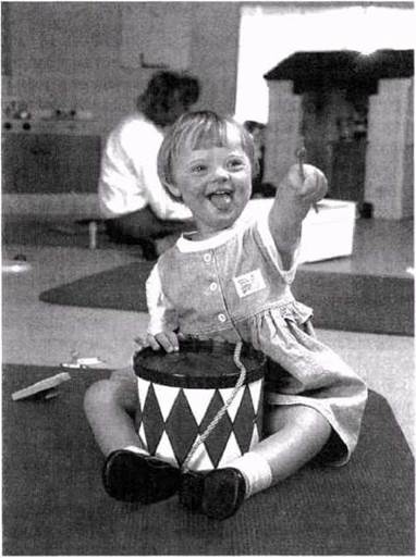
Fig. 25.27. A girl showing Characteristic Features of Down's syndrome: slightly misaligned eyes, a rounded HEAD, and a flat nasal bridge.
Explanation
In 96% of cases, the cause of Down's syndrome is the nondisjunction of chromosome 21 during the anaphase of meiosis. Theoretically, this can happen during The formation of either sperm or egg Cells, but the latter occurs much more frequently (see below regarding METABOLISM/18.html">The Influence of maternal age). Approximately 70% of nondisjunction events occur in meiosis I and 30% in meiosis II. In meiosis I, nondisjunction involves entire chromosomes, whereas in meiosis II, it involves sister chromatids. The outcome is the same: two chromosomes or two chromatids end up in one daughter cell, while the other receives none (Fig. 24.31). This process should theoretically produce an equal number of cells with monosomy 21; however, this condition, like all cases of monosomy, is lethal to the fetus at early Selection/3.html">Stages of development.
In approximately 3–4% of cases, Down's syndrome is the result of a translocation (Section 24.9.2). Chromosome 21 becomes attached to chromosome 14 or, less commonly, to chromosome 22.
The Influence of Maternal Age
The probability of giving birth to a child with Down's syndrome depends heavily on the mother's age (Fig. 25.28), whereas no correlation with paternal age has been found. At age 20, the risk is 1 in 2,000; at 30, it is 1 in 900; at 40, it is 1 in 100; and at 44, it reaches 1 in 40. When these data are plotted as a curve on a logarithmic scale, it becomes apparent that the slope of the curve steepens progressively with age (Fig. 25.28). This is likely because a woman's oocytes are formed during embryonic development and subsequently mature one by one each month once reproductive age is reached. In contrast, new sperm cells are continuously produced in men from Puberty until death.
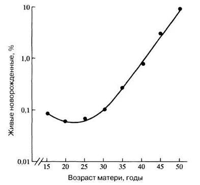
Fig. 25.28. The Effect of maternal age on the probability of having a child with Down syndrome. Note that the vertical axis is logarithmic.
Other examples of autosomal trisomies
In most cases, autosomal trisomies prove to be lethal, resulting in miscarriage in the Cytology/cytology/16.html">Early stages of fetal development. In cases of trisomy involving very small chromosomes, namely chromosomes 13 and 18, a lethal effect may be avoided; however, the children born present with numerous defects, including mental retardation. Typically, they fail to thrive normally and die within the first three months of life.
Last update: 06/08/2026
Editorial and Educational Adaptation: This material has been compiled based on the primary/original source text. The project team performed an editorial review, corrected technical inaccuracies, structured sections, and adapted the content for an educational format.
What was processed:
- elimination of formatting defects (OCR errors, structural breaks, corrupted characters);
- editorial organization of content;
- standardization of terminology in accordance with academic sources;
- verification of factual statements against the original source text.
All mentions of the author, publication year, and origin of the primary text have been preserved in accordance with the source.