BIOLOGY Volume 3 - A Guide to General Biology - 2004
25. APPLIED GENETICS
25.1. Bacterial Genetic Engineering
25.1.2. Stage 1. Obtaining a Copy of the Required Gene
This is the most challenging part of the process. For example, The Human Genome (which is all the DNA within a Cell) contains approximately 3 billion NUCLEOTIDES and = 100,000 genes (according to recent data ≈ 30,000). A typical Gene is several thousand Base Pairs long, making even locating a specific gene quite difficult. Three main Methods are used to obtain a gene copy:
1) using Reverse Transcriptase to produce a gene copy based on its mRNA template;
2) synthesizing an artificial gene;
3) using the "shotgun" method, which involves cleaving DNA with restriction Enzymes (Restriction Endonucleases) and searching for the fragment containing the desired gene.
We will illustrate the techniques of Introduction/32.html">Genetic Engineering first by using the first two methods, which lead directly to the goal—namely, isolating the required gene. We will then examine the third method.
Using Reverse Transcriptase
Despite the fact that a diploid cell contains only two copies of each gene (one on the chromosome inherited from the mother, and another on the chromosome from the father), an actively functioning gene typically produces thousands of complementary mRNA molecules (sec. 23.8). It is usually known in which Cells a given gene is active. For example, the gene encoding Insulin is active in the pancreatic β-cells. Retroviruses contain an enzyme that can synthesize a complementary DNA copy from an RNA molecule. Producing DNA from an RNA template is the reverse of normal METABOLISM/31.html">Transcription, where RNA is synthesized on a DNA template; hence, the enzyme is called reverse transcriptase. It has become a valuable tool in genetic engineering. (Retroviruses themselves use this enzyme to convert their own RNA genome into DNA capable of infecting new cells; sec. 2.4.5.) If the cells in which the gene of interest is active are known, isolating mRNA from them is straightforward. Once this is accomplished, the mRNA is converted by reverse transcriptase into a DNA copy of the desired gene (Fig. 25.1). DNA obtained in this manner is called complementary DNA, or cDNA, regardless of whether it is single-stranded or double-stranded.
Class="center">
Fig. 25.1. Genetic engineering. A schematic diagram of the Procedure developed for Gene cloning. Details are explained in the text.
Gene Synthesis
The base sequence of a gene can be determined either directly or derived from the Amino Acid Sequence of the protein encoded by that gene. A gene can then be constructed from nucleotides (recall that each base is part of a single nucleotide) by joining them together in the correct order. Currently, only short genes can be constructed this way, but with methodological improvements, the synthesis of any gene will become a routine procedure. This method has been used to obtain the proinsulin and Somatostatin genes. Somatostatin (also known as Growth Hormone-inhibiting factor) is a protein hormone consisting of just 14 Amino Acids.
The "Shotgun" Method - Using Restriction Enzymes
This primary method of gene isolation was developed in the late 1960s and early 1970s. Its emergence was made possible by the discovery of enzymes called restriction endonucleases or restriction enzymes. These enzymes, found in Bacteria, are capable of cutting DNA; for instance, they cleave any viral DNA invading a bacterial cell, thereby restricting the reproduction of Viruses within The Cell. Different species of bacteria produce various restriction endonucleases. Each of them cleaves the nucleic acid (hence "nuclease") at strictly defined points ("endo" means that the enzyme cleaves the molecule from within rather than attacking it from the ends). The enzyme recognizes a specific base sequence and interacts precisely with it. The Cleavage sites are called restriction sites. To date, over 2,000 restriction enzymes active against 230 different sequences have been isolated. The bacterium protects its own DNA by attaching methyl groups to specific bases within the restriction sites.
The name of each enzyme is derived from the bacterium from which it is isolated (Fig. 25.2). Note that the target restriction sequence is often six bases long and palindromic, meaning it reads the same in both directions. When examining Fig. 25.2, keep in mind that the two complementary DNA strands are read in opposite directions. Some restriction enzymes make staggered cuts. As a result of their action, DNA fragments with protruding single-stranded ends are formed (e.g., EcoRI, Fig. 25.2). Such ends are called "sticky ends"; they are used to rejoin DNA fragments. They essentially stick together through The formation of Hydrogen Bonds with complementary sticky ends of other DNA molecules cut by the same restriction enzyme (Fig. 25.1). For instance, the widely used EcoRI restriction enzyme produces a TTAA sticky end. Other restriction enzymes generate blunt ends. An example of this is HindII (Fig. 25.2). Using restriction enzymes, the DNA of any Organism can be fragmented. The length of the resulting restriction fragments depends on the type of restriction endonuclease used and the Location OF THE base sequences recognized by that enzyme (Fig. 25.3).
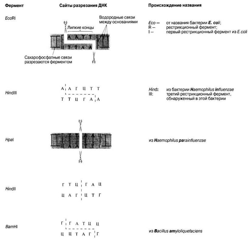
Fig. 25.2. Some commonly used restriction enzymes. EcoRI, HindIII, and BamHI make zig-zag (staggered) cuts in DNA, leaving "sticky ends". A sticky end produced by EcoRI, for example, can join with another sticky end produced by EcoRI. HindII and HpaI leave blunt ends. Schematics for EcoRI and HpaI are presented in greater detail.
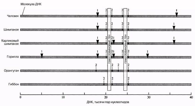
Fig. 25.3. The Use of restriction enzymes to cut DNA and produce restriction fragments of varying lengths. In this figure, a DNA segment containing two Hemoglobin Genes from different primates was treated with two restriction enzymes. Enzyme 1 is indicated by  and enzyme 2 by ↓. Enzyme 1 cuts human DNA into two fragments, while enzyme 2 cuts it into five fragments. The lengths of these fragments, as well as those obtained from double Digestion with both enzymes, make it possible to map the relative positions of the cuts (restriction sites) to one another. Based on these data, a "restriction map" is constructed, as shown in the diagram. The more restriction enzymes used to build the map, the more detailed it becomes. Note that the closer the evolutionary relationship between species, the more similar their DNA and the arrangement of their restriction sites. [Diagram based on Fig. 7–4, p. 294, Molecular Biology of the Cell, 3rd ed., B. Alberts et al. (1994) Garland.]
It is known that every nucleotide in DNA carries a negatively charged phosphate group, meaning that DNA fragments of different lengths carry proportional charges. These differences can be used to separate DNA fragments in an electric field via gel Electrophoresis (Fig. 25.4). As the name implies, the method involves the use of an agarose gel (for very large fragments) or a polyacrylamide gel (for smaller fragments). Since DNA is colorless, THE POSITION OF a particular fragment in the gel after electrophoresis is revealed either by staining or by using radioactively labeled DNA and conducting autoradiography, during which a photographic film is placed over the gel. The radioactive radiation exposes the film precisely where the DNA is located.
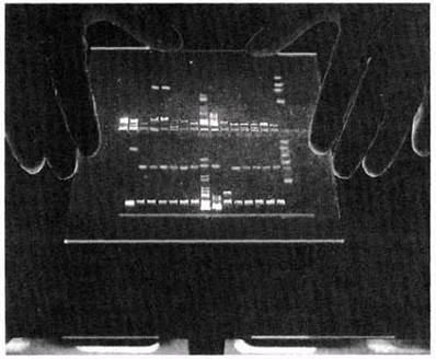
Fig. 25.4. Gel electrophoresis used to separate DNA fragments of varying lengths. The fragments are generated by cutting DNA with one or more restriction enzymes. The largest fragments migrate the slowest because it is much harder for them to pass through the Pores in the gel. In the photograph, they are located near the sample wells in the gel.
When donor DNA is digested with restriction enzymes, it is assumed that one of the fragments will randomly contain a complete copy of the target gene and nothing else. This non-specific gene isolation approach is known as shotgun cloning. The most challenging task when using this method is identifying the fragment that carries the desired gene, a problem discussed in Section 25.1.3.
Interrupted genes
Using reverse transcriptase or chemical gene synthesis has an advantage over the shotgun method because the resulting gene is not "interrupted". Interrupted genes contain one or more DNA regions called introns that do not code for The amino acid sequence of a protein. The function of introns remains unclear. It is only known that if a eukaryotic gene containing introns is introduced into a bacterium, the bacterium will synthesize a useless protein because bacteria lack the enzymes required to remove introns from mRNA. How introns are removed from mRNA is illustrated in Fig. 25.5.
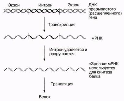
Fig. 25.5. Transcription and Translation of an intron-containing gene. The regions flanking the intron are called exons. Genes may contain many introns. Proteins are encoded solely by exons.
Last update: 06/08/2026
Editorial and Educational Adaptation: This material has been compiled based on the primary/original source text. The project team performed an editorial review, corrected technical inaccuracies, structured sections, and adapted the content for an educational format.
What was processed:
- elimination of formatting defects (OCR errors, structural breaks, corrupted characters);
- editorial organization of content;
- standardization of terminology in accordance with academic sources;
- verification of factual statements against the original source text.
All mentions of the author, publication year, and origin of the primary text have been preserved in accordance with the source.